Marchiafava-Bigmani Disease in Relation to Pure Alcohol Consumption
To the Editor: Marchiafava-Bignami disease (MBD) is a rare complication of alcoholism defined by the presence of symmetrical demyelization and necrosis of the corpus callosum, causing neuropsychiatric manifestations. It was first described by Marchiafava and Bignami, who identified it in the autopsies of chronic wine drinkers who presented with seizures and coma.1 Nowadays, the diagnosis is supported by the association of suggestive clinical data and brain magnetic resonance (MRI).2 In this report, we describe a case of acute MBD after a period of massive pure alcohol consumption.
Case Report
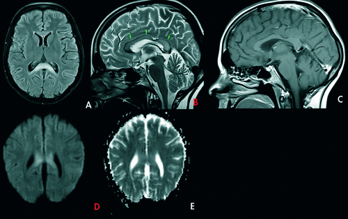
A 30-year-old homeless woman with a past history of postpartum depression 2 years ago, presented in an acute confusional state. She had been binge-drinking in the last year. Four weeks preceding admission, she started massive daily intake of commercially available pure alcohol (70%–90%), approximately 250–300 ml /day, or 175–270 mg/day, leading to emergency hospitalization on two occasions. She was aggressive, and showed massive generalized hypertonicity, with hyperactive reflexes. The cranial nerves were intact. No signs of meningeal irritation was noticed. Laboratory results (cerebrospinal fluid assay, metabolic panel) were normal, with exception of hypochromic macrocytic anemia (hemoglobin: 98 g/L, mean globular volume: 110 fL) and mild elevation of hepatic enzymes (alanine transaminase: 66 units/L; aspartate transaminase: 55 units/L). The electroencephalogram showed lentification of the cerebral activity. Brain cranial tomography (CT) was unremarkable. Brain MRI on T2W, DWI, and FLAIR images showed hypersignal of corpus callosum, without water restriction. On T1W and apparent diffusion coefficient (ADC), the lesion appeared hypointense (Figure 1). Based on the history of alcohol abuse in association with the imaging findings, the diagnosis of MBD was made. Despite supportive measures, intravenous vitamin B complex, and methylprednisolone, she died a week later.
Discussion
MBD is mostly reported in malnourished middle-aged men with a history of severe chronic alcoholism.1 Brain MRI is the most sensitive method of diagnosis. It shows areas of hypointensity in T1, hyperintensity on T2, and FLAIR signal in the corpus callosum.1,2 Our patient exhibited the typical imaging findings of MBD. The differential diagnosis with Wernicke encephalopathy (WE), which is a more common alcohol complication, is mandatory, but WE and MBD can occur simultaneously . The absence of ophthalmoplegia or abnormal signal of the mammillary bodies, thalamus, or brainstem, made the diagnosis of WE unlikely.1,3 A wide diversity of alcohol beverages have been implicated in MBD.1 The patient was taking massive amounts of pure alcohol in the previous month. There is no specific therapy for MBD. Cessation of alcohol is mandatory. Treatment with thiamine and vitamin B complex can be effective,1,4 and is recommended under the presumption of an underlying vitamin deficiency.1,5 Mortality in MBD is high (21%), and those who survive tend to have severe dementia or are bedridden.1,2,5 In conclusion, we report a case of MBD unique for the association with pure alcohol consumption, which has never been described before.
1 : Diseases of the nervous system due to nutritional deficiency: Marchiafava-Bignami disease (primary degeneration of the corpus callosum), in Adams and Victor's Principles of Neurology. Ropper AHSamuels M. New York, McGraw-Hill, 2005, pp 998–999Google Scholar
2 : Clinicoradiologic subtypes of Marchiafava-Bignami disease. J Neurol 2004; 251:1050–1059Crossref, Medline, Google Scholar
3 : A non-alcoholic Japanese patient with Wernicke’s encephalopathy and Marchiafava-Bignami disease. Clin Neuropathol 1984; 3:231–236Medline, Google Scholar
4 : A case of Marchiafava-Bignami disease: complete recovery with thiamine. J Neuropsychiatry Clin Neurosci 2011; 23:E28Link, Google Scholar
5 : [Marchiafava-Bignami disease]. Rev Neurol 1999; 28:519–523Medline, Google Scholar