Depressive Disorder After Pontine Ischemic Stroke: Clinicoradiologic Correlates
To the Editor: It is widely known that the occurrence of poststroke mood disorders, especially depression, is one of the most frequent complications of stroke.1 Studies evaluating the association of focal lesions with depressive disorder have shown that lesions involving the frontal lobe, caudate, or putamen were more likely to lead to depression than comparable isolated lesions of the brainstem.2,3 To the best of our knowledge, there are no studies in the literature showing the metabolic correlates of poststroke depression that is secondary to an isolated brainstem infarction.
Case Report
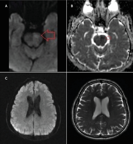
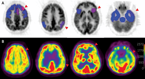
A 61-year-old man experienced depressive symptoms after he sustained a left-sided pontine stroke 1 month earlier. The patient described having severe physical fatigue, moderate insomnia, and suicidal thoughts in the last few weeks. He felt sad and lost practically all interest in doing things, which led him to retreat from daily work activities. The patient was cooperative, alert, and fully oriented during the psychiatric examination. He displayed a depressed mood state. His speech was slowed, and he spoke in a moderate depressive voice. The patient scored 24 points on the original Beck Depression Inventory, which indicated moderate depression. The neurological examination result was normal except for right-sided mild motor weakness (4+/5). A diffusion-weighted MRI scan of the brain performed on the admission day showed an acute infarct on the left pontine area and a corresponding apparent diffusion coefficient (Figure 1, A and B). By contrast, reduced glucose uptake in [18F]fluorodeoxyglucose and positron emission tomography scans also involved the left frontal and temporoparietal cortices (Figure 2), whereas there were no abnormalities in the left frontal and temporoparietal cortical regions on MRI (Figure 1C).

FIGURE 1. Diffusion-Weighted MRI Scana
a(A) The diffusion-weighted MRI scan displays hyperintensity on the left pontine area, which corresponds with an area of decreased apparent diffusion coefficient located in the (B) left pons and theoretically represents the area of cytotoxic edema and acute infarct (red arrows). (C) On diffusion-weighted MRI and fluid-attenuated inversion recovery sequences, respectively, there were no abnormalities in the left frontal and temporoparietal cortical regions that might explain the apparent hypometabolism revealed by [18F]fluorodeoxyglucose and positron emission tomography imaging.

FIGURE 2. [18F]fluorodeoxyglucose and Positron Emission Tomography Imagesa
a(A) NeuroQ software (version 3.5; Syntermed, Inc., Atlanta, GA) was used to process the (B) raw [18F]fluorodeoxyglucose and positron emission tomography data. The average pixel values in standardized regions of interest were automatically calculated. Area/whole brain ratios were compared with the normal values in the database. Significant hypometabolism is seen on the left frontal parietal and temporal cortical areas.
Discussion
The brainstem has traditionally been associated with vital functions, reflexive emotional reactions, and motor performance. Although the neuronal activity within the brainstem with motor areas of the cerebral cortex was found to correlate highly with parameters of movement, the role of the brainstem in the development of depression is still unclear. In the case of our patient, functional abnormalities did not entirely parallel morphological changes and were also found in the frontal, temporal, and parietal regions, which appeared to be rather unaffected on MRI. This indicates that the reduced glucose uptake observed in the respective cortical regions may reflect secondary deficits as a result of diminished functions of emotional circuits involving the brainstem. This was suggested by previous studies showing that with a depressive mood state, increases in limbic-paralimbic blood flow and decreases in neocortical regions were identified in depression.4 Our findings suggest that the functional integrity of pathways linking the cortex and brainstem may be integral to the normal regulation of mood; these results also indicate a relationship between the brainstem noradrenergic system and depression. This type of quantitative analysis can provide that information, unlike a subjective radiologic evaluation limited with MRI. A greater understanding of the functional activity of the underlying regions affected by poststroke depression may help to not only provide insight into the specific networks involved, but it may also support the finding that disruption of the neurochemistry of noradrenergic transmission plays an important role in the pathophysiology of major depression.5
1 : Post-stroke depression, antidepressant treatment and rehabilitation results. A case-control study. Cerebrovasc Dis 2001; 12:264–271Crossref, Medline, Google Scholar
2 : Poststroke depression: a review. Can J Psychiatry 2010; 55:341–349Crossref, Medline, Google Scholar
3 : Early depressive symptoms after stroke: neuropsychological correlates and lesion characteristics. J Neurol Sci 2005; 228:27–33Crossref, Medline, Google Scholar
4 : Reciprocal limbic-cortical function and negative mood: converging PET findings in depression and normal sadness. Am J Psychiatry 1999; 156:675–682Medline, Google Scholar
5 : Reduced levels of norepinephrine transporters in the locus coeruleus in major depression. J Neurosci 1997; 17:8451–8458Crossref, Medline, Google Scholar


