Voltage-Gated, Potassium-Channel Antibody-Associated Limbic Encephalitis Presenting as Acute Psychosis
Case Presentation
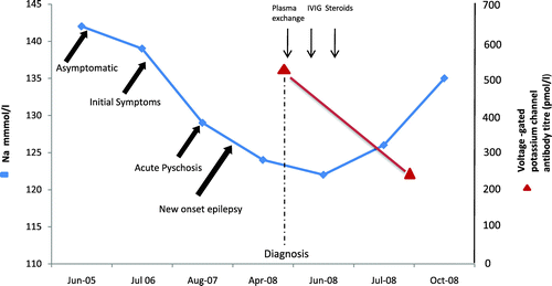
A 58-year-old man was admitted to the hospital in April 2008 with increasing frequency of seizures, confusion, slurred speech, ataxia, and falls. He had a previous history of excess alcohol consumption, depression, and deliberate self-harm. The patient's depressive symptoms began in August 2006 and were associated with episodes of amnesia and confusion. A year later (August 2007) the patient was admitted with an episode of severe self-harm. This was particularly disturbing, as the patient had self-inflicted multiple stab wounds with a knife to his neck, chest, abdomen, and upper limbs. Computed tomography (CT) scan of the chest and abdomen revealed a left pneumothorax, left pleural effusion, and free gas in the peritoneal cavity. Urgent laparotomy showed multiple anterior abdominal wall lacerations but no intra-abdominal organ injury. A chest drain was inserted to drain the left hemo-pneumothorax. Upper limb injuries included multiple right flexor tendon injuries and a deep laceration to the left biceps tendon, all of which required surgical repair. A psychiatric assessment at this stage concluded that the patient was suffering with paranoid delusions and auditory and visual hallucinations; he was subsequently diagnosed with paranoid schizophrenia. He was started on olanzapine, which resulted in some improvement of his psychotic symptoms. Over the next 2 months, his cognitive functioning deteriorated significantly, with progressive amnesia, personality change in the form of aggression, and new-onset epilepsy. Comparison of his previous Mini-Mental State Exam scores showed a rapid decline, from 26/30 to 8/30 in 2 months. His epilepsy worsened, and the seizures became both bizarre and resistant to multiple anticonvulsants. On this admission, blood investigations were all normal, apart from low serum sodium. The hyponatremia (ranging from 122 mmol/liter to 126 mmol/liter) in retrospect had persisted over 10 months (Figure 1). Plasma and urine osmolality suggested this to be due to the syndrome of inappropriate ADH secretion (SIADH). A brain MRI showed nonspecific areas of high signal. CSF analysis and an EEG were normal. Investigations for neurological infections, malignancy (CT scans of the chest, abdomen, and pelvis, and tumor markers), onco-neuronal, autoimmune, and thyroid antibodies were all negative, but serum voltage-gated, potassium-channel antibodies (VGKC-Abs) were elevated, at 506 pmol/liter (normal range: 0–100 pmol/liter). A diagnosis of limbic encephalitis (LE) was made on the basis of these findings. The patient was treated with plasma exchange, followed by intravenous immunoglobulins and corticosteroids. A repeat VGKC antibody titer 3 months later showed a reduction to 253 pmol/liter, which coincided with improvement in serum sodium levels, as well as improvement in the patient's seizure frequency and aggressive behavior. There was minor improvement in his cognitive functioning, although it is likely that the patient had suffered with autoimmune (VGKC antibody-associated) LE for nearly 2 years, which may have caused permanent neuronal damage irreversible by immunotherapy.

Discussion
Neuropsychiatric symptoms form a key part of VGKC antibody-associated LE and are often the presenting feature of this condition.1–5 Autoimmune-mediated damage to limbic structures result in amnesia, cognitive decline, and epilepsy. Acute psychosis of this severity has not been previously reported, and it highlights the diversity of this condition's clinical spectrum. It is difficult to determine precisely when our patient developed this condition, but we assume it was at least 12 months before the episode of acute psychosis. The initial episodes (August 2006) of confusion, memory loss, and depressive symptoms could well have been the first manifestations of the illness. The patient's psychotic episode, which resulted in extreme self-mutilation (August 2007) was deemed to be primarily due to a psychiatric cause. The possibility of an organic cause was not considered—ultimately delaying the diagnosis. It was only several months later that he began to deteriorate more rapidly, with symptomatic memory loss, cognitive decline, and seizures that led to the diagnosis of LE nearly 2 years after his initial symptoms. Epilepsy and hyponatremia appear to be important features of this condition's clinical syndrome. Seizures appear to develop later in the disease and are often both diverse and difficult to treat.1–5 Hyponatremia is well described in several case series and reports of LE; the cause is thought to be SIADH, as was the case in our patient. In nearly all cases, the hyponatremia is resistant to standard treatment such as fluid restriction, but corrects after treatment with immunoglobulins and/or plasma exchange.1,4,5 In this case, the onset of hyponatremia coincided with the onset of his clinical deterioration, suggesting a temporal relationship and a potential marker for disease activity. (The association between serum sodium measurements and clinical symptoms over a 3-year period is shown in Figure 1). VGKC antibody-associated encephalitis is a subacute, degenerative neurological condition potentially reversible with immunotherapy. Neuropsychiatric symptoms in the presence of persistent hyponatremia and seizures are key presenting features that should alert the clinician to the possibility of autoimmune LE. Acute psychosis is a rare and unusual presentation, but if associated with other clinical features suggestive of LE, testing for VGKC antibodies should be performed.
1. : Potassium channel antibody-associated encephalopathy: a potentially immunotherapy-responsive form of limbic encephalitis. Brain 2004; 127:701–712Crossref, Medline, Google Scholar
2. : Voltage-gated potassium channel antibody-mediated syndromes: a spectrum of clinical manifestations. Rev Neurol Dis 2008; 5:65–72Medline, Google Scholar
3. : Voltage-gated potassium channel antibody-associated encephalitis with basal ganglia lesions. Neurology 2006; 66:1780–1781Crossref, Medline, Google Scholar
4. : A case of voltage-gated potassium channel antibody-related limbic encephalitis. Nat Clin Pract Neurol 2006; 2:339–343Crossref, Medline, Google Scholar
5. : Psychiatric presentation of voltage-gated potassium channel antibody-associated encephalopathy. Br J Psychiatry 2006; 189:182–183Crossref, Medline, Google Scholar


