Acute, Non-Lethal Marchiafava-Bignami Disease With Reversible Posterior Leucoencephalopathy and Split-Brain Syndrome
Case Report
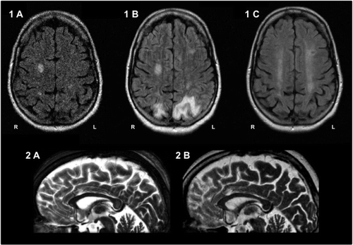
A 44-year-old woman with history of alcohol abuse and depression was admitted, unconscious, to our hospital with a Glasgow Coma Score of 3, SpO2 of 50% in the periphery, and blood alcohol level of 2.11%. Neurological examination revealed bilaterally positive Babinski sign; initial cranial computerized tomography and CSF examination showed no pathologies. Laboratory examinations showed normochromic macrocytic anemia, hyponatremia, hypokalemia, and hypocalcaemia, and massively elevated liver and pancreatic enzymes. An intensive-care treatment with blood transfusion and high-dose catecholamine application became necessary, and a high-dose intravenous infusion of Vitamin-B-complex was initiated. Within the first days, the patient progressively regained consciousness, while spontaneous verbal production and repetition were absent. Further neurological examination revealed ataxia, astasia, and abasia. Neuropsychological testing showed an interhemispheric disconnection syndrome, with persisting expressive speech disability but preserved comprehension, right–left confusion, ideomotor apraxia, agraphia, acalculia, pseudoneglect, and memory deficits. Cranial MRI (cMRI) 1 week after admission demonstrated a lesion in the splenium of the corpus callosum (CC), as well as bilateral lesions in the centrum semiovale (CS; Figure 1[A], Figure 2[A]). Two weeks after admission, generalized epileptic seizures emerged, and neurological examination confirmed cortical blindness. Follow-up cMRI (Day 16) showed lesions in both occipital lobes, whereas the lesion in the CC could no longer be detected (Figure 1[B], Figure 2[B]). Another cMRI 1 month after admission showed a complete involution of the occipital lesions (Figure 1[C]). Two months later, the patient was able to walk without help. Cortical blindness had completely resolved, whereas the split-brain syndrome persisted.

Discussion
MBD typically results in a progressive demyelinization and cystic necrosis of the CC. Also, the pathogenic process may extend into the extracallosal neighboring white matter, and even subcortical and cortical regions.3 Our patient showed an acute onset of MBD with a lesion in the splenium of the CC and bilateral lesions in CS. After intensive-care treatment, the course of the disease was not lethal; however, neuropsychological testing revealed a split-brain syndrome, normally found in the chronic form of MBD. Follow-up cMRI studies showed complete regression of the callosal lesion and incomplete regression of the lesions in the centrum semiovale. Also, cMRI at Day 16 revealed a reversible posterior leucoencephalopathy correlated with symptomatic epileptic seizures and cortical blindness, a symptom not typically seen in MBD. Whereas there have been reports of MBD with favorable outcome,4 our patient is one of few with nonlethal acute MBD, providing evidence that the acute form of MBD may also have a prolonged course with continuous gradual improvement, as well an association with reversible posterior leucoencephalopathy. Immediate intensive-care treatment with symptomatic therapy and thiamine infusion may be critical in the prevention of a lethal outcome.
1. : Marchiafava-Bignami disease. Neurology 1978; 28:290–294Crossref, Medline, Google Scholar
2. : Signs of interhemispheric disconnection in Marchiafava-Bignami disease. Arch Neurol 1977; 34:254Crossref, Medline, Google Scholar
3. : Cortical involvement in Marchiafava-Bignami disease. Am J Neuroradiol 2005; 26:670–673Medline, Google Scholar
4. : Marchiafava-Bignami disease with widespread lesions and complete recovery. Am J Neuroradiol 2010; 31:1506–1507Crossref, Medline, Google Scholar


