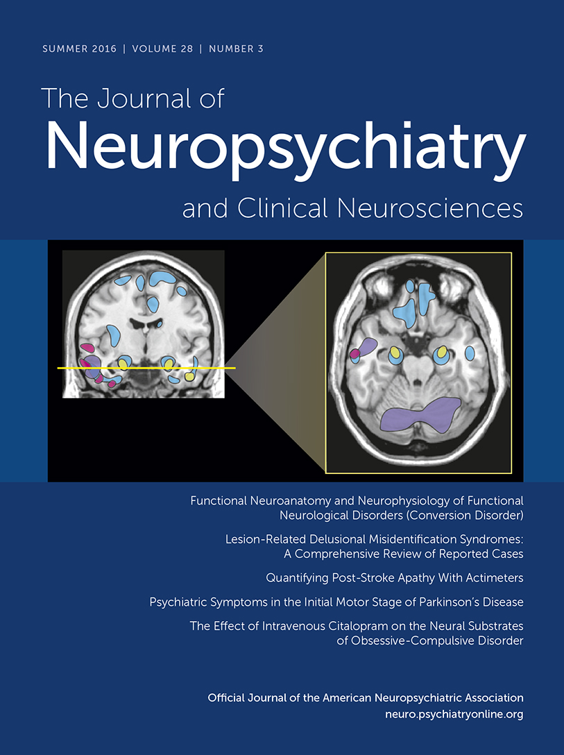
Lesion-Related Delusional Misidentification Syndromes: A Comprehensive Review of Reported Cases
Abstract
Delusional misidentification syndromes (DMSs) are persistent delusions of hyper- or hypofamiliarity for meaningful persons and places in one's environment. This study set to determine the clinical course, neuroanatomical localization, neuropsychological abnormalities, and delusional content in patients with DMSs occurring after focal neurological injuries. Sixty-one patients were identified: 28 with hypofamiliar delusions, 27 with hyperfamiliar delusions, and, most surprisingly, six patients with both hypo- and hyperfamiliar delusions. Recognition is often delayed by months from the time of injury, and the delusions are self-limited. Patients with DMSs had right hemisphere (92%) injuries (specifically right frontal injuries in 63%), prominent memory impairment (73%), and multiple concurrent DMSs (29%).
Delusional misidentification syndromes (DMSs) are a fascinating group of disorders involving a fixed, false belief about the identity of persons, places, and objects in one’s environment. These include delusions in which a familiar person or place is misperceived as being an unfamiliar imposter (Capgras delusion) or delusions in which a strange person or place is misperceived as being familiar (Fregoli delusion; Table 1). Although DMSs were initially described in psychiatric patients, such presentations have been increasingly reported in patients with focal neurological lesions.1
| Type of Delusion | Person | Place | Self | Object |
|---|---|---|---|---|
| Hypofamiliarity | Capgras | Capgras place | Cotard, mirror misidentification | Capgras object |
| Example | Misidentify mother as an imposter | Misidentify home as replica | Believe that one is dead (Cotard) | Misidentify car as replica |
| Hyperfamiliarity | Fregoli, intermetamorphosis | Reduplicative paramnesia | Subjective double | n/a |
| Example | Misidentify doctor as dead mother (Fregoli) | Believe home reduplicated inside hospital | Believe another version of oneself exists |
TABLE 1. Delusional Misidentification Syndromes
DMSs are often conceptualized as a “two-hit” process, in which one deficit produces an abnormal perception, whereas a second deficit leads to the persistence of that abnormal perception as a delusion impervious to reasoning.2,3 Although right frontal dysfunction is associated with the pathological persistence of delusional beliefs after neurological injuries,4–7 hypotheses regarding the genesis of abnormal belief content in DMSs remain controversial. In Capgras syndrome, it has been proposed that the delusion results from a disconnection between implicit and explicit facial processing8; from a disconnection between visual facial perceptual areas and limbic structures9; from dysfunction in representations of the intentions, beliefs, and feelings of others10; or from dysfunction in self-relational processes.11
The goal of this study was to systematically examine the clinical, neuroanatomical, neuropsychological, and delusional characteristics of all reported cases of DMSs after acute neurological injury. These analyses will help guide and constrain theories on the genesis of delusions in DMSs and will provide prognostic information about the typical clinical course of DMSs.
Methods
Case Selection
We performed a PubMed search to identify all reported cases of lesion-related DMSs through Dec. 31, 2014. We used an inclusive search strategy to identify both (a) the DMS component (using the terms “delusional misidentification” OR “Capgras” OR “Fregoli” OR “intermetamorphosis” OR “subjective doubles” OR “reduplicative paramnesia” OR “Cotard”) and (b) the acute neurological injury (using the search terms “MRI” OR “CT” OR “SPECT” OR “PET” OR “stroke” OR “hemorrhage” OR “bleed” OR “trauma” OR “TBI”). Inclusion criteria included occurrence of a DMS after an acute neurological injury (e.g., a stroke, hemorrhage, or trauma), with neuroanatomical imaging confirming such injury. We defined a DMS as a delusion (fixed, false belief impervious to reasoning) involving inappropriate feelings of familiarity or unfamiliarity toward a person, place, or object of personal significance to the patient. We excluded patients in whom sufficient clinical information was lacking to determine whether the nature of the delusion was consistent with a DMS, patients with ongoing delusions or psychotic symptoms from a primary psychiatric disease or delirium, and cases in which only functional neuroimaging (e.g., positron emission tomography or single photon emission computed tomography) was available. To assess the possibility that premorbid psychiatric disease contributes to delusion maintenance, we did not exclude patients with chronic but stable psychiatric disease in whom new delusions developed in the setting of an acute neurological injury.
Data Analysis and Classification
Demographic information (age, gender), type of injury, time to onset, and time to resolution were collected from reported cases. In addition, delusions were classified according to the direction of impaired familiarity (hypofamiliar, hyperfamiliar, or both), object category (person, place, object, self, event), presence of multiple DMSs, and presence of other delusions. Neuroimaging results were classified according to laterality (right, left, bilateral) as well as gross localization (frontal, temporal, parietal, occipital, basal ganglia, thalamus). When images were included, these were used to confirm reported localization. Neuropsychological evaluations were assessed to determine the presence or absence of deficits within the domains of memory, executive function, language, and visuospatial function. In addition, we noted instances of comorbid prosopagnosia, neglect, limb denial, or confabulation given the potential relevance of these syndromes in some theoretical models of DMSs.
Statistical Analyses
All statistical analyses were performed using the STATA software package (version 14.0; StataCorp LP, College Station, TX), with p<0.05 for significance. Fisher’s exact tests were used to determine the significance of neuroanatomical localization (e.g., right versus left hemisphere, frontal lobe versus other lobes), neuropsychological deficits (e.g., memory versus executive versus language versus visuospatial), and whether there were significant differences between patients with hypofamiliar and hyperfamiliar delusions.
We did not have a method for directly assessing the severity of DMS. However, the duration of the delusions could possibly relate to delusion severity. Therefore, in an exploratory analysis, we used linear regression to determine whether the delusion duration is related to unilateral versus bilateral hemispheric lesions, lesions within specific cortical lobes, or specific neuropsychological deficits. Variables significantly predicting delusion duration (p<0.05) were included in the final regression model. Age and etiology of the lesion were additionally controlled for in all regression analyses.
Results
Patient Demographic and Clinical Information
Our search yielded 507 articles, from which 61 patients were identified as meeting our inclusion/exclusion criteria (Table 2, Supplemental Table 1). The mean age was 52 years and 67% of patients were men. The most common etiologies included ischemic stroke (thrombotic and embolic), hemorrhagic stroke (nontraumatic deep, lobar, and subdural hemorrhages), and trauma (contusions and/or traumatic hemorrhages). Other etiologies included infections (toxoplasmosis and neurocysticercosis), tumors and tumor resections, and hemiplegic migraine with cerebral edema. There was often a delay between injury and recognition of the DMS (median 60 days). The duration of delusions varied widely, with a median duration of 42 days before resolution. Longer reported durations were often attributable to recurrence of delusions in the setting of medical or psychiatric decompensation. Premorbid psychiatric disease was rare (7%).
| Etiology | Demographics | Clinical Features | |||||||
|---|---|---|---|---|---|---|---|---|---|
| Type of Delusion | Ischemic | Hemorrhagic | Trauma | Other | Psychiatric Diagnosis | Men | Age, Yearsb | Days to Onsetc | Days to Resolutiond |
| Total (N=61) | 24 (39) | 12 (20) | 17 (28) | 8 (13) | 4 (7) | 41 (67) | 52 (56) | 130 (60) | 220 (42) |
| Hypofamiliar (N=28) | 11 (39) | 6 (21) | 7 (25) | 4 (14) | 2 (7) | 18 (64) | 50 (52) | 136 (105) | 343 (60) |
| Hyperfamiliar (N=27) | 10 (37) | 6 (22) | 9 (33) | 2 (7) | 1 (4) | 19 (70) | 55 (60.5) | 116 (30) | 74 (30) |
| Both (N=6) | 3 (50) | 0 (0) | 1 (17) | 2 (33) | 1 (17) | 4 (67) | 47 (47) | 205 (205) | 231 (81) |
TABLE 2. Demographic and Clinical Featuresa
Neuroimaging Analysis
Neuroimaging results are reported in Table 3. Fisher’s exact tests showed that right hemisphere lesions (92% of cases) were more common than left hemisphere lesions (p<0.001), and right frontal lesions (63%) were more common than lesions within other right hemisphere lobes or the left hemisphere (p<0.001). Left hemispheric lesions were more commonly seen in patients with hypofamiliar delusions than those with hyperfamiliar delusions (p=0.01). A linear regression showed that, after controlling for age and etiology, bilateral hemispherical involvement resulted in significantly longer delusion duration, compared with unilateral hemispheric lesions (beta=374, 95% confidence interval=9–740, p=0.05). A linear regression showed no significant relationship between delusion duration and involvement of specific cortical lobes (p=n.s.).
| Hemisphere | Right | Left | ||||||||||||
|---|---|---|---|---|---|---|---|---|---|---|---|---|---|---|
| Type of Delusion | Right | Left | Bilateral | Right/Bilateral | Frontal | Parietal | Temporal | Occipital | Basal Ganglia/Thalamus | Frontal | Parietal | Temporal | Occipital | Basal Ganglia/Thalamus |
| Total (N=61) | 44 (72) | 5 (8) | 12 (20) | 56 (92)* | 39 (63)* | 20 (32) | 18 (29) | 7 (11) | 7 (8) | 13 (21) | 1 (2) | 5 (8) | 1 (2) | 4 (6) |
| Hypofamiliar (N=28) | 16 (57) | 3 (11) | 9 (32) | 25 (89) | 18 (64) | 8 (29) | 5 (18) | 3 (11) | 3 (11) | 9 (32) | 0 (0) | 4 (14) | 1 (4) | 2 (7) |
| Hyperfamiliar (N=27) | 24 (89) | 0 (0) | 3 (11) | 27 (100) | 19 (70) | 10 (37) | 10 (37) | 3 (11) | 1 (4) | 2 (7) | 1 (4) | 1 (4) | 0 (0) | 0 (0) |
| Both (N=6) | 4 (67) | 2 (33) | 0 (0) | 4 (67) | 2 (33) | 2 (33) | 3 (50) | 1 (17) | 1 (17) | 2 (33) | 0 (0) | 0 (0) | 0 (0) | 2 (33) |
TABLE 3. Neuroimaging Abnormalitiesa
Neuropsychological Analysis
Fifty-six patients (92%) reported having cognitive and neurological testing; only 31 (50%) patients underwent formal neuropsychological testing. Deficits in memory, executive function, and visuospatial impairment were common in patients with DMSs (Table 4), with memory impairment being the most common (p<0.001). Patients with hyperfamiliar delusions were more likely to have executive dysfunction compared with patients with hypofamiliar delusions (p=0.01). A linear regression showed that, after controlling for age and etiology, the presence of confabulations significantly predicted longer delusion duration (beta=682, 95% confidence interval=329–1036, p<0.001).
| Type of Delusion | Facial Recognition | Memory | Executive Function | Visual | Language | Attentional Neglect | Confabulatory Memory Disorder | Somatoparaphrenia |
|---|---|---|---|---|---|---|---|---|
| Total (N=56) | 9 (16) | 41 (73)* | 29 (52) | 21 (38) | 3 (5) | 18 (32) | 7 (13) | 1 (2) |
| Hypofamiliar (N=24) | 5 (21) | 18 (75) | 8 (33) | 8 (33) | 3 (13) | 5 (21) | 5 (21) | 1 (4) |
| Hyperfamiliar (N=27) | 3 (11) | 19 (70) | 18 (67)** | 11 (41) | 0 (0) | 11 (41) | 2 (7) | 0 (0) |
| Both N=5 | 1 (20) | 4 (80) | 3 (60) | 2 (40) | 0 (0) | 2 (40) | 0 (0) | 0 (0) |
TABLE 4. Neuropsychological Deficitsa
Delusion Characteristics
Delusion characteristics are reported in Table 5. Contrary to classification schemes dividing DMSs based on hypo- or hyperfamiliar delusions, we found that the presence of both hypo- and hyperfamiliar delusions was not rare (10%). A total of 29% of patients had DMSs toward multiple classes of objects (persons, places, self, object, event). In addition, 11% of patients with DMSs had other non-DMS delusions as well, mostly consisting of persecutory paranoid ideation.
| Type of Delusion | Person | Place | Object | Self | Event | Multiple |
|---|---|---|---|---|---|---|
| Total (N=61) | 33 (50) | 38 (58) | 2 (3) | 9 (14) | 6 (9) | 19 (29) |
| Hypofamiliar (N=28) | 17 (59) | 14 (45) | 1 (3) | 4 (13) | 0 (0) | 8 (26) |
| Hyperfamiliar (N=27) | 11 (41) | 20 (74) | 0 (0) | 1 (4) | 4 (15) | 7 (26) |
| Both (N=6) | 5 (83) | 4 (67) | 1 (17) | 4 (67) | 2 (33) | 4 (67) |
TABLE 5. Misidentificationa
Discussion
Main Findings
The main findings of our study are as follows. First, recognition of lesion-related DMS is often delayed from the acute neurological injury. Second, most cases are self-limited, although the presence of bilateral lesions or confabulations predicts longer delusion duration. Third, injuries commonly involve damage to the right hemisphere and frontal lobes. Fourth, patients show a range of impairments on formal neuropsychological testing, most notably memory impairment. Finally, delusions of hypo- and hyperfamiliarity and delusions for persons and places often coexist within the same patient, suggesting a shared mechanism. Taken together, these results provide valuable information for practitioners encountering these disorders in their patients and offer potential insights into the underlying neural mechanisms leading to abnormal belief content in DMSs.
Clinical Features of DMSs
We found that, in general, there was a delay to diagnosis of DMSs, although this varied widely from immediate presentation to several years after the injury. There are several possible explanations for this finding. First, the delusions may have been masked by other neurological symptoms (neglect, apathy, or other neuropsychiatric symptoms; level of arousal) or misattributed to general confusion or delirium. Practitioners in rehabilitation and/or outpatient settings may also be more familiar with these delusions than clinicians providing acute care. It is also possible that these disorders are not a direct effect of the lesion but arise later as a consequence of maladaptive plasticity occurring after the injury. A similar mechanism is thought to explain the delayed onset of tremor and some movement disorders that can occur up to months after an acute injury.12
We did not find that premorbid psychiatric disease, dementia, or advanced age were common in our identified patients, suggesting that these predisposing conditions are not necessary for DMSs to occur. Although we attempted to omit cases of obvious concurrent delirium, we cannot definitively exclude the possibility that comorbidities common in hospitalized stroke patients, including sleep disturbances, unreported infections, or electrolyte abnormalities, contributed to the onset and/or maintenance of DMSs.
Neuroanatomical Lesions in DMSs
A previous review of reported cases of DMSs with available data on neuroanatomical lesions found that 28 (97%) of 29 cases involved the right frontal lobe.4 Our larger group of 61 patients confirms this general pattern of right-sided and frontal lesions being more common, with posterior lesions and left-sided lesions being significantly less common. Our study, however, showed a wider distribution of lesions, with only 63% involving the right frontal lobe. This difference may be attributable to our broader search strategy and inclusion criteria. In addition, the previous study included cases of limb denial, which might have a more precise localization to the right frontal lobe and is not universally considered a DMS.
Alternatively, the fact that many lesions causing the DMS were located outside of the right frontal lobe may suggest that DMSs result from a disturbance of a distributed brain network, focused in the right frontal lobe but involving regions outside of this location. Supporting the hypothesis that the network effects of a lesion may correlate with delusion formation better than a specific anatomical localization, a recent functional magnetic resonance imaging study of a patient with Capgras delusion showed reduced activation in regions distant from his right frontal lesion (left precuneus and superior temporal sulcus).13 Involvement of a distributed network would also explain our finding of longer delusion duration associated with bilateral hemispheric lesions compared with unilateral lesions, despite not finding any association between delusion duration and a specific lobar location of the lesion.
Neuropsychological Deficits in DMS
We found that neuropsychological deficits were reported across multiple cognitive domains, including memory, executive functioning, and visuospatial processing. Memory was statistically more likely to be impaired than other cognitive domains, consistent with previous claims that memory may play an important role in the development of these delusions.14,15 The presence of confabulations predicted significantly longer delusion duration, again supporting the importance of memory impairment in DMSs. Language deficits were notably absent, likely owing to the right hemispherical dominance of the lesions. However, it is possible that language impairment from left-sided lesions would potentially mask DMSs in these patients.
We found evidence for abnormal face processing in patients with DMSs, although the inconsistency in methods of assessing facial perception prevents strong conclusions about specific impairments in these patients. According to the model of Ellis and Young,8 damage to the ventral visual pathway leads to prosopagnosia (impaired conscious face recognition), whereas damage to the dorsal visual areas leads to an impaired sense of familiarity for known faces. Other popular models of facial perception have instead proposed an “extended-facial system” that complement the “core” facial perceptual areas within the ventral stream; these extended areas mediate multiple aspects of facial perception, including retrieval of pertinent semantic information, dynamic facial features involved in theory of mind processes, and retrieval of salient autobiographical and personality features.16 It is possible that abnormalities in these higher-order features of facial recognition lead to the specific deficits in facial perception that form the substrate for some types of DMSs.
Delusional Content in DMSs
One surprising finding from our analysis is that many of our patients had multiple delusional misidentifications simultaneously. Some patients, for example, experienced DMSs for both a person (e.g., their spouse) as well as a place (e.g., their home). More strikingly, other patients experienced delusions of both hyperfamiliarity and hypofamiliarity, such as believing that one’s wife was an imposter and that one’s new doctor was a deceased father in disguise. This finding contradicts theories that different DMSs, such as Capgras and Fregoli syndromes, result from distinct mechanisms. For example, theories that view Capgras syndrome as a disconnection between facial recognition areas and limbic regions8,9,17,18 or regions involved in theory of mind10 do not explain how delusions of hyperfamiliarity or delusions for place could exist in the same patient. Our findings therefore identify an uncommonly recognized feature of DMSs that should be accounted for in a comprehensive theoretical explanation of how these delusions occur.
One potential explanation is that DMSs result from an inability to link perceptions of external stimuli in the environment with internally generated autobiographical memories.19 Hypofamiliar delusions would result when externally perceived objects do not trigger appropriate internal autobiographical memories. In the case of Capgras delusion, the failure to retrieve the appropriate autobiographical memories associated with a family member would lead to the erroneous belief that the family member is an imposter. Hyperfamiliar delusions would result when internally generated autobiographical memories pertaining to a particular person or location are not appropriately suppressed by the conflicting external percept. In Fregoli delusion, for example, internally generated autobiographical memories about a loved one are not appropriately constrained when meeting one’s physician, leading to the erroneous interpretation that one’s doctor is a family member in disguise. This proposed explanation predicts disruption in distributed neural networks and specific deficits in autobiographical memory, and both of these predictions are supported by the results from this study.
Limitations
By virtue of its retrospective design, our study has several limitations. First, in the review of published cases, clinical information was limited to details provided in individual reports. Because these reports were not focused on the specific characteristics reported here, information was often incomplete. Further prospective studies are clearly needed in order to confirm our findings. In addition, our neuroimaging analysis involved gross anatomical localization given the heterogeneity of reporting, and cases often did not include images for more quantitative lesion-overlap analysis. Such analyses would be useful in further clarifying the mechanisms of such delusions. In a similar fashion, neuropsychological testing was heterogeneous, limiting the strength of conclusions regarding specific associated neuropsychological deficits.
Conclusions
DMSs are fascinating, bizarre, and poorly understood, but the study of patients in whom these disorders arise from acute neurological injuries can provide invaluable information about the nature of these delusions. Our study shows that the recognition of these delusions is typically delayed and that most are self-limited; in addition, the presence of bilateral hemispheric lesions and confabulations predicts longer delusion duration. Our results confirm localization to the right hemisphere and frontal lobe, but identified lesions in other locations suggest that more distributed networks may be important in delusion formation. Furthermore, our finding of significant memory impairment, out of proportion to other neuropsychological functions, suggests that amnesia might be particularly important in the formation of these delusions. Finally, our results demonstrate less recognized features of DMSs, such as the common co-occurrence in the same patient of hypo- and hyperfamiliar delusions, or delusions for persons and for places, which suggests that the various types of DMSs exist on a spectrum.
1 : Delusional misidentifications and duplications: right brain lesions, left brain delusions. Neurology 2009; 72:80–87Crossref, Medline, Google Scholar
2 : The neuropsychology of delusions. Ann N Y Acad Sci 2010; 1191:16–26Crossref, Medline, Google Scholar
3 : Cognitive neuropsychiatry and delusional belief. Q J Exp Psychol (Hove) 2007; 60:1041–1062Crossref, Medline, Google Scholar
4 : The Lost Self: Pathologies of the Brain and Identity. New York, Oxford University Press, 2005Crossref, Google Scholar
5 : Delusional thoughts and regional frontal/temporal cortex metabolism in Alzheimer’s disease. Am J Psychiatry 2003; 160:341–349Crossref, Medline, Google Scholar
6 : Neurobiology of delusions, memory, and insight in Alzheimer disease. Am J Geriatr Psychiatry 2014; 22:1346–1355Crossref, Medline, Google Scholar
7 : Grey matter atrophy in mild cognitive impairment/early Alzheimer disease associated with delusions: a voxel-based morphometry study. Curr Alzheimer Res 2015; 12:165–172Crossref, Medline, Google Scholar
8 : Accounting for delusional misidentifications. Br J Psychiatry 1990; 157:239–248Crossref, Medline, Google Scholar
9 : Capgras syndrome: a novel probe for understanding the neural representation of the identity and familiarity of persons. Proc Biol Sci 1997; 264:437–444Crossref, Medline, Google Scholar
10 : The misidentification syndromes as mindreading disorders. Cogn Neuropsychiatry 2010; 15:233–260Crossref, Medline, Google Scholar
11 : Neuropathologies of the self: clinical and anatomical features. Conscious Cogn 2011; 20:75–81Crossref, Medline, Google Scholar
12 : Movement disorders in cerebrovascular disease. Lancet Neurol 2013; 12:597–608Crossref, Medline, Google Scholar
13 : When a loved one feels unfamiliar: a case study on the neural basis of Capgras delusion. Cortex 2014; 52:75–85Crossref, Medline, Google Scholar
14 : On reduplicative paramnesia. Brain 1903; 26:242–267Crossref, Google Scholar
15 : Reduplicative paramnesia: a disconnection syndrome of memory. Cortex 1982; 18:23–35Crossref, Medline, Google Scholar
16 : Neural systems for recognition of familiar faces. Neuropsychologia 2007; 45:32–41Crossref, Medline, Google Scholar
17 : Capgras delusion: an interactionist model. Conscious Cogn 2008; 17:863–876Crossref, Medline, Google Scholar
18 : In what sense ‘familiar’? Examining experiential differences within pathologies of facial recognition. Conscious Cogn 2009; 18:628–638Crossref, Medline, Google Scholar
19 : “Cat-gras” delusion: a unique misidentification syndrome and a novel explanation. Neurocase (Epub ahead of print, Jan. 14, 2016)Medline, Google Scholar