Hippocampal Volume Increased After Cognitive Behavioral Therapy in a Patient With Social Anxiety Disorder: A Case Report
To the Editor: Functional imaging studies have revealed that patients with social anxiety disorder have hyperactivity of the amygdala and surrounding areas, including the hippocampus.1 However, the number of structural brain imaging studies is limited. Irle et al.2 reported that social anxiety disorder patients had reduced amygdala (13%, only male patients) and hippocampus (8%) size and that a smaller right hippocampus is associated with more severe symptoms.
Although some studies detected that the hippocampus/amygdala increase after effective treatments in patients with depression or other anxiety disorders,3,4 there are no morphological studies in patients with social anxiety disorder.
Here we describe a case in which the hippocampus increased after cognitive-behavioral therapy (CBT).
Case
A 36-year-old woman with an 8-year history of social anxiety disorder received 12 weeks of group CBT.
Scores for the Liebowitz Social Anxiety Scale,5,6 Social Phobia Scale, and Social Interaction Anxiety Scale7,8 were obtained before, immediately after, and 3 months after CBT.
We obtained MRI data before and 3 months after CBT using a 1.5-T MRI system (Gyoro Scan Intera; Philips Medical Systems, Best, the Netherlands). The scanning parameters for the three-dimensional T1-weighted turbo field echo sequences were as follows: echo time, 3.9 msec; repetition time, 8.5 msec; flip angle, 15°; 256 × 256 matrices; field of view, 25 cm; voxel size, 1.0 × 1.0 × 1.0 mm; and slice thickness, 1.0 mm.
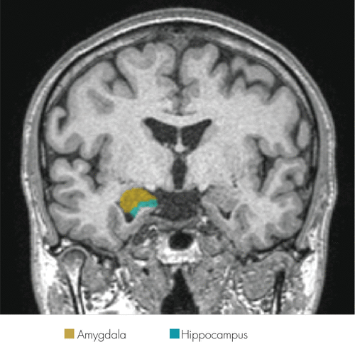
The hippocampus/amygdala were traced manually according to the method used in a previous study9 (Figure 1): an operator blind to subject status traced them twice, and a comparison of the two tracings was made. When the operator used another MRI dataset (N=24), the interrater/intrarater reliability of the hippocampus was 0.89/0.90 and that of the amygdala was 0.80/0.61.

FIGURE 1. Manual Tracing of Hippocampal and Amygdala Volumes
The patient gave written consent, and this study was approved by the Ethics Committee of Nagoya City University.
The volume of the right hippocampus increased from 3417.51 to 3890.50 mm3, and the Liebowitz Social Anxiety Scale scores decreased from 109 to 70 and , Social Interaction Anxiety Scale/Social Phobia Scale scores decreased from 72/43 to 42/19. Clinically, her avoidant behaviors were decreased. The volume of the right amygdala was decreased, and the volume of the left hippocampus/amygdala did not change (Table 1).
| Before CBT | 3 Months After CBT | Volumetric Change | |
|---|---|---|---|
| LSAS total score | 109 | 70 | |
| SIAS total score | 72 | 42 | |
| SPS total score | 43 | 19 | |
| Left hippocampus (mm3) | 3028.51 | 3007.00 | –0.71% |
| Right hippocampus (mm3) | 3417.51 | 3890.50 | 13.84% |
| Left amygdala (mm3) | 1026.50 | 995.50 | –3.02% |
| Right amygdala (mm3) | 1239.00 | 1097.00 | –11.46% |
TABLE 1. Clinical Data and Hippocampal/Amygdala Volume Change
Discussion
We described our experience with a patient with social anxiety disorder whose right hippocampal volume increased 3 months after successful CBT.
Previous studies have shown that prolonged stress and a high level of glucocorticoids reduce hippocampus/amygdala volumes and that effective treatment alleviates the situation in major depressive disorders. However, in the case of anxiety disorders, the mechanism of volume reduction is not clear.
Irle et al.2 reported that, in female patients with social anxiety disorder, only the hippocampi were smaller than those of healthy control subjects, and the small right hippocampi had an association with symptom severity. Our finding was supported by this study.
However, the reason for the reduction in the right amygdala is unclear and could be becasue of our unskilled tracing technique.
Conclusion
Although larger sample studies are needed, we expect that the right hippocampal volume may have an association with symptom or treatment response in patients with social anxiety disorder.
1 : Functional neuroimaging of anxiety: a meta-analysis of emotional processing in PTSD, social anxiety disorder, and specific phobia. Am J Psychiatry 2007; 164:1476–1488Crossref, Medline, Google Scholar
2 : Reduced amygdalar and hippocampal size in adults with generalized social phobia. J Psychiatry Neurosci 2010; 35:126–131Crossref, Medline, Google Scholar
3 : [Evaluation study of clinical and neurobiological efficacy of EMDR in patients suffering from post-traumatic stress disorder]. Riv Psichiatr 2012; 47(Suppl):12–15Medline, Google Scholar
4 : Long-term treatment with paroxetine increases verbal declarative memory and hippocampal volume in posttraumatic stress disorder. Biol Psychiatry 2003; 54:693–702Crossref, Medline, Google Scholar
5 : Social phobia. Mod Probl Pharmacopsychiatry 1987; 22:141–173Crossref, Medline, Google Scholar
6 : Reliability and validity of the Japanese version of the Liebowitz Social Anxiety Scale. Seishin Igaku (Clinical Psychiatry) 2002; 44:1077–1084Google Scholar
7 : Development and validation of measures of social phobia scrutiny fear and social interaction anxiety. Behav Res Ther 1998; 36:455–470Crossref, Medline, Google Scholar
8 : Development and validation of the Japanese version of Social Phobia Scale and Social Interaction Anxiety Scale. Jpn J Psychosom Med 2004; 44:841–850Google Scholar
9 : Left hippocampal volume inversely correlates with enhanced emotional memory in healthy middle-aged women. J Neuropsychiatry Clin Neurosci 2007; 19:335–338Link, Google Scholar


