Late Onset of Psychotic Symptoms in a Patient With Cavum Septum Pellucidum and Cavum Vergae
Case Report
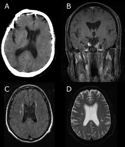
A 64-year-old man had first onset of visual hallucinations at age 60, associated with fear of being hurt; these were controlled by antipsychotic drugs. Six months ago, he developed gait disorder. Recently, he presented with atypical schizophrenia-like psychosis, with disorders of speech and movement. CT (Figure 1[A]) and MRI (Figure 1 [B, C, D]) showed an infarctive lesion in the right internal capsule and bilateral enlarged lateral ventricles, along with CSP and CV. He scored 14 on the Mini-Mental State Exam (MMSE). He was managed medically with aspirin, statins, neuroprotective drugs, and potent antipsychotic drugs. His mental status was normalized; his motor akinesia was diminished, as well as other symptoms ameliorated after the use of more potent antipsycotic drug and drugs that enhanced cerebral blood flow.

[A] CT scan showing slight low density in the right internal capsule and bilateral enlargement of occipital portion of the lateral ventricle along with cavum septum pellucidum and cavem vergae. [B] Coronal MRI showing the CSP and CV. [C] MRI T1WI and [D] MRI T2WI showing high-intensity lesion in the right internal capsule and enlargement of lateral ventricles with presence of cavum septum pellucidum and cavum vergae.
Discussion
The prevalence of CSP and CV is about 0 · 15%, and affects men more than women.(3) The size of normal-variant cavum is approximately 1 mm–4 mm. CSP is considered abnormally large if it is ≥ 6 mm in size. Enlarged CSP is often seen in psychiatric patients(1,2) and considered as evidence of disturbed midline development of the brain, and particularly of the limbic system.(5) The symptomatic cava could be managed medically, but the medications only reduce signs and symptoms; also, there is development of tolerance and quick recurrence. Surgery should be an option in management because through surgery these midline cysts could be incised and stop further enlargement. In the case reported, the patient first presented mild psychotic symptoms, which remained stable for few years, followed by exacerbation, which indicates the progressive nature of the disease. Global atrophy of the brain might be responsible for the gradual enlargement of cavities, and atrophy of limbic and paralimbic structures might have enhanced psychotic symptoms. Miyamori et al.(4) concluded that the thickening of the septum pellucidum and the lowering of the ventricular pressure were the two main factors in developing the pathological cava. Structural and functional deficit in limbic and paralimbic regions usually cause associative and integrative problems, such as distorted interpretations of reality. Also, brain midline abnormalities and the age factor synergistically might have accelerated cognitive decline; hence, he had a lower score on the MMSE. Paresis of the left limbs was caused by an infarctive lesion in the right internal capsule, which improved after treatment. The possibility of drug tolerance could not be excluded since the patient's symptoms gradually recovered after medical management. Surgical treatment, if necessary, was left for future management.
Conclusion
Enlarged CSP and CV are a neurodevelopmental anomaly and are associated with psychotic behavior. Asymptomatic CSP and CV should be screened yearly. Symptomatic CSP and CV should be managed medically or surgically to prevent further enlargement and psychosis.
1. : Cavum septum pellucidum in schizophrenia: clinical and neuropsychological correlates. Psychiatry Res 2007; 154:147–155Crossref, Medline, Google Scholar
2. : MRI study of cavum septi pellucidi in schizophrenia, affective disorder, and schizotypal personality disorder. Am J Psychiatry 1998; 155:509–515Crossref, Medline, Google Scholar
3. : Clinical correlates of septum pellucidum cavities: an unusual association with psychosis. Psychol Med 1985; 15:43–54Crossref, Medline, Google Scholar
4. : Expanded cavum septi pellucidi and cavum vergae associated with behavioral symptoms relieved by a stereotactic procedure: case report. Surg Neurol 1995; 44:471–475Crossref, Medline, Google Scholar
5. : The septum pellucidum: normal and abnormal. Am J Neuroradiol 1989; 10:989–1005Medline, Google Scholar


