Choroid Plexus Papilloma Presenting as Schizophrenia: A Case Report
To the Editor: Although the definite etiology of schizophrenia is not known, there have been reports of intracranial tumors presenting with schizophrenia symptoms. In this report, a first such case, we present a patient who had classical symptoms of schizophrenia, who was, on investigation, found to have a choroid plexus papilloma. This case emphasizes the need for imaging in patients having signs of organicity and supports the concept of cognitive dysmetria in patients with schizophrenia.
The etiology of schizophrenia remains obscure. Recent studies and reviews have implicated genetic and environmental risk factors as causative agents, along with the observation of neuro-immunologic, white-matter disconnectivity, and neurophysiological correlations as some of the consistent associations with schizophrenia.1,2 However, there have been occasional reports that an intracranial lesion may present with symptoms of schizophrenia.3 In this case report, we present a patient who presented with symptoms of schizophrenia, who later on investigation was diagnosed as having choroid plexus papilloma.
Case Report:
"Mr. V," a 38-year-old man, presented to our hospital in July 2008 with a 2-year history of slowness in activity, lethargy, and loss of interest in previously pleasurable activities. For the previous 6 months, these symptoms were associated with delusions of persecution, reference, and third-person auditory hallucinations in the absence of any mood or anxiety symptoms. These symptoms were associated with intermittent headache, predominantly occipital, and a 15-day history of intermittent gait imbalance. He had no history of any substance abuse and had deterioration in the socio-occupational functioning in the previous 6 months.
Physical examination revealed presence of right-sided cerebellar signs, and mental status examination revealed impaired prosody of speech and corroborated the delusion of persecution, reference, and third-person auditory hallucinations. The diagnosis of schizophrenia was considered.
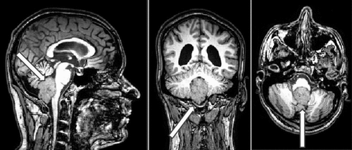
In view of the late onset of psychosis, unusual clinical presentation, and positive cerebellar signs, magnetic resonance imaging (MRI) of the brain was done. MRI showed a uniformly enhancing, space-occupying lesion in the fourth ventricle, extending through the foramen of Magendie into the cisterna magna (Figure 1). After this finding on the imaging studies, he was referred to a neurosurgeon and was taken to surgery with a plan for complete excision of the growth on the 10th day of the admission. During the operative process, the growth was seen in the cistern magna, extending up into the fourth ventricle through the foramen of Magendie and extending downward into the foramen magnum and covering the lower part of the cerebellum. Postoperatively, he had rapid relief from headache and gait imbalance. Histopathological examination of the growth confirmed the diagnosis of choroid plexus papilloma. The patient was evaluated on the 5th postoperative day, during which time he did not have any psychotic symptoms but was noted to have a few anxiety symptoms. When he was interviewed again after 2 weeks, he had completely improved, with neither psychotic nor anxiety symptoms.

Discussion
To the best of our knowledge, this is the first report of an adult with choroid plexus papilloma presenting with schizophrenia symptoms. Choroid plexus is an epithelial–endothelial convolute in the ventricular system of the brain, which is neuroectodermal in origin.4 The major functions of the choroid plexus are secretion of cerebrospinal fluid (CSF) and maintenance of an intact blood–brain barrier.5 Choroid plexus papilloma is a benign tumor arising out of the choroid plexus. The most common sites for papilloma to appear (in decreasing order) are lateral ventricles, and fourth ventricles, followed by third ventricles. The symptoms include increased intracranial tension, focal neurologic deficits, cranial nerve palsies, seizures, and coma.6 One report of choroid plexus papilloma in a 9-year-old boy who presented with history suggestive of psychosis has been reported in the literature.3
The patient in our report presented with classical schizophrenia symptoms. Detailed physical examination and evaluation with imaging threw light into the underlying choroid plexus papilloma, removal of which resulted in complete remission of symptoms without the need for any antipsychotic medication.
We would like to highlight another observation in this case: the relation of the tumor to the cerebellum and its mass effect. Aberration in the circuit connecting the cerebellum and the cerebral cortex, the corticocerebral–thalamo–cortical circuit (CCTCC), leads to impaired modulation of functioning, which is termed “cognitive dysmetria”.7–9 Evidence suggests cognitive dysmetria as the basis for varied symptoms of schizophrenia, such as hallucinations, delusions, disorganization, and negative symptoms.8
Thus, schizophrenia patients presenting with an unusual clinical profile, with subtle indicators of organicity should undergo imaging of brain to rule out organic cause. This case also supports the possible role of cerebellar dysfunction in the pathogenesis of schizophrenia.
1 : Schizophrenia, “just the facts:” what we know in 2008, 2: epidemiology and etiology. Schizophr Res 2008; 102:1–18Crossref, Medline, Google Scholar
2 : Schizophrenia, “just the facts:” what we know in 2008, 3: neurobiology. Schizophr Res 2008; 106:89–107Crossref, Medline, Google Scholar
3 : Third ventricular choroid plexus papilloma with psychosis: case report. J Neurosurg 1997; 87:103–105Crossref, Medline, Google Scholar
4 : Choroid plexus: biology and pathology. Acta Neuropathol 2010; 119:75–88Crossref, Medline, Google Scholar
5 : The choroid plexus in the rise, fall, and repair of the brain. Bioessays 2005; 27:262–274Crossref, Medline, Google Scholar
6 : Choroid plexus tumors in children: significance of stromal invasion. Neurosurgery 2001; 48:303–309Medline, Google Scholar
7 : Structural cerebellar abnormalities in antipsychotic-naive schizophrenia: evidence for cognitive dysmetria. Indian Journal of Psychological Medicine 2008; 30:14–20Crossref, Google Scholar
8 : “Cognitive dysmetria” as an integrative theory of schizophrenia: a dysfunction in cortical–subcortical–cerebellar circuitry? Schizophr Bull 1998; 24:203–218Crossref, Medline, Google Scholar
9 : Prefrontal cortex projections to the basilar pons in rhesus monkey: implications for the cerebellar contribution to higher function. Neurosci Lett 1995; 199:175–178Crossref, Medline, Google Scholar


