Wernicke’s Encephalopathy in Two Different Clinical Settings: One After Whipple Surgery and the Other Due to Alcohol Abuse
To the Editor: Wernicke’s encephalopathy (WE) is an acute neuropsychiatric disorder because of thiamine (vitamin B1) deficiency, which is characterized by a classic triad of mental status changes, ocular motor abnormalities, and ataxia.1 WE is a life-threatening condition, and its prognosis depends on prompt diagnosis followed by immediate supplementation of thiamine. Here, we report two cases of WE with different etiologies, one after Whipple surgery because of pancreatic carcinoma and the other associated with chronic alcoholism.
Case Reports
Case #1
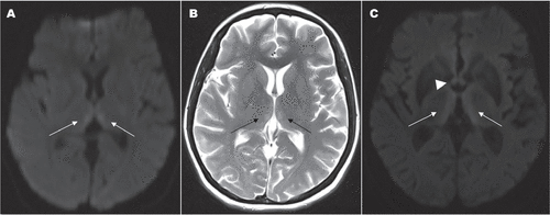
A 38-year-old woman was diagnosed with carcinoma of the head of the pancreas and underwent pancreaticoduodenectomy (Whipple procedure). Ten days after the initial surgery, she underwent a second surgery because of the obstruction of jejunum. She received total parenteral nutrition without thiamine supplementation. On the sixth day after the second operation, on her neurological examination upbeat nystagmus in the primary position of gaze and bilateral horizontal gaze-evoked nystagmus with an oblique direction because of upbeat nystagmus were detected. Bilateral dysmetria, truncal, and gait ataxia were also observed. Brain MRI revealed increased signal on T2 and diffusion weighted image (DWI) sequences in the medial regions of bilateral thalami (Figure 1, A and B). Two days after the beginning of intravenous administration of thiamine, the patient was oriented and she could stand by herself. On the 15th day, she was discharged with intact neurological examination, except for minimal residual vertical nystagmus.

FIGURE 1. Symmetrical high-intensity signals are seen in the medial thalami in diffusion-weighted image (white arrows) [A] and T2-weighted axial MRI (black arrows) [B] of case 1. Note symmetrical signal-intensity alterations in the mammillary bodies (arrowhead) and medial thalami (white arrows) in DWI of case 2 [C].
Case #2
A 59-year-old man was admitted to the emergency department because of fluctuation in the level of consciousness and hallucinations for 2 days. He had a history of excessive alcohol consumption for the last 30 years. On neurological examination, he was confused, disoriented, and confabulating. He had also dysartric speech, horizontal nystagmus, minimal horizontal conjugate gaze palsy, and prominent truncal ataxia. DWI MRI showed symmetrical hyperintense lesions in the medial regions of bilateral thalami (Figure 1, C). His routine laboratory testing was normal. In the context of severe alcoholism, this clinical presentation was highly suggestive of WE, and immediate parenteral thiamine replacement was started. After initiation of thiamine therapy, significant improvement was observed within 48 hours. He was discharged at the end of the 2-week follow-up, with mild memory impairment and ataxia.
Discussion
WE is a well-defined thiamine-deficiency syndrome. Confusional state, oculomotor manifestations, and gait ataxia are the clinical triad of WE. However, many other neurological symptoms and signs may also appear, especially if there is a delay in treatment.1,2 WE is mostly seen in association with chronic alcoholism in developed countries. Other predisposing factors and clinical conditions associated to WE are the gastrointestinal disease and surgery, malignancy, hyperemesis gravidarum, malnutrition, severe infections, and dialysis.1,3 Severe vomiting and parenteral nutrition without thiamine supplementation may contribute to the early occurrence of WE after the operation, as observed in our patient.
The diagnosis of WE remains mostly clinical, and the best aid for a correct diagnosis is still clinical suspicion in the appropriate settings. Alcoholism, and persistent vomiting after gastrointestinal surgeries should be considered alarming.
The most frequently affected regions are the medial thalamus and periventricular region of the third ventricle (80%), followed by periaqueductal area (59%) and mammillary bodies (45%).4
If WE is suspected, prompt treatment with parenteral thiamine should be administered until there is no further improvement in signs and symptoms.5 Prophylactic thiamine supplementation should be considered in patients with alcoholism and malnutrition.
In conclusion, early replacement of thiamine reverses the neurological signs of WE and prevents permanent brain damage, it should be supplemented rapidly and adequately in all cases at risk of malnutrition, such as alcoholism and gastrointestinal disorders.
1 : Wernicke’s encephalopathy: new clinical settings and recent advances in diagnosis and management. Lancet Neurol 2007; 6:442–455Crossref, Medline, Google Scholar
2 : Wernicke encephalopathy after sleeve gastrectomy. Isr Med Assoc J 2012; 14:708–709Medline, Google Scholar
3 : Wernicke’s encephalopathy induced by total parental nutrition. Nutr Hosp 2010; 25:1034–1036Medline, Google Scholar
4 : MR imaging findings in 56 patients with Wernicke encephalopathy: nonalcoholics may differ from alcoholics. AJNR Am J Neuroradiol 2009; 30:171–176Crossref, Medline, Google Scholar
5 :


