Thalamic and Thalamic Reticular Nucleus Abnormalities in a Patient With Chromosome 22q11.2 Deletion Syndrome
To the Editor: Chromosome 22q11.2 deletion syndrome (22q11.2DS) is one of the common human deletion syndromes, with developmental delay, cognitive impairment, schizophreniform psychosis, and several physical symptoms. Although several neuroradiological findings, mainly about brain structural abnormalities, have been detected, little is known about the neural basis of cognitive and psychiatric impairment. In this report, we present rare findings on ethyl cysteinate dimer (ECD) single photon emission CT (SPECT) and MRI revealing abnormalities of the thalamus and thalamic reticular nucleus that may be associated with a psychiatric disorder.
Case Report
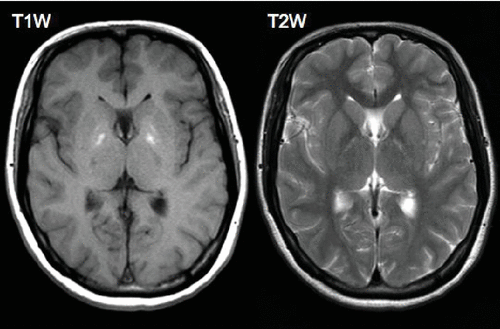
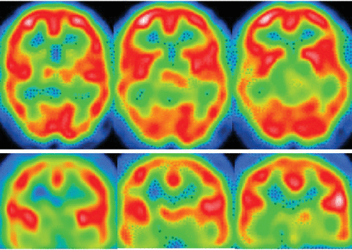
A 24-year-old woman developed occipital headache. She was diagnosed as having 22q11.2DS on genetic testing at 7 years of age. Her medical history was comprised of a tetralogy of Fallot and a congenital defect of the right carotid artery. She had been attending the department of psychiatry for schizophreniform psychosis for a long time. On examination, she presented with headache and anxiety. No abnormality was found on neurologic examination except for mild mental retardation. Brain MRI data showed linear high-intensity lesions on T2-weighted imaging and low-intensity lesions on T1-weighted imaging in the outer margin of the thalamus adjacent to the internal capsule (i.e., thalamic reticular nucleus, bilaterally) (Figure 1). SPECT with Tc-99m ECD revealed hypoperfusion in the bilateral thalamus (Figure 2). She was diagnosed as having a tension-type headache.

FIGURE 1. Brain MRI Data Showed High-Intensity Lesions in the Bilateral Thalamic Renticular Nucleus on T2-Weighted Imaging and Low-Intensity Lesions on T1-Weighted Imaging
T1-weighted imaging showed mild high intensity in the bilateral globus pallidus, indicating calcification.

FIGURE 2. SPECT With Tc-99m ECD Revealed Hypoperfusion in the Bilateral Thalamus
Discussion
Prominent findings in neuroimaging studies on 22q11.2DS patients are reduction of overall brain volume, midline defects, structural alterations of the cerebellum and frontal lobes, white matter abnormalities, decreased gray matter volumes in parietal and temporal areas, enlarged lateral ventricles, and increased basal ganglia volumes. In relation to visuospatial and other cognitive impairments, reduction volume of the superior portion of the thalamus, particularly in the medial and posterior regions including the pulvinar nucleus,1 and atypical development of the cortical connective patterns using diffusion tensor image data2 have been detected. Among functional imaging studies, only one 123I-IMZ-SPECT study revealing a gyrus abnormality and related epileptic evidence has been reported.3 This is the first report of neuroradiological findings revealing a thalamic abnormality on SPECT and a thalamic reticular nucleus abnormality on MRI. From the anatomical and functional aspects, whole thalamic hypoperfusion on SPECT may be influenced by the thalamic reticular nucleus abnormality because of the connectivity.
The thalamus and cerebral cortices are the most important regions for the generation of consciousness, behavioral performance, cognition, emotion, and thought. The thalamic reticular nucleus, a shell-shaped nucleus located between the thalamus and the cortices that has a connective role between the three principal neurons that comprise the corticothalamic, thalamocortical, and reticulothalamic systems, plays central roles in information integration and in the generation of conscious experience. Pathogenically, it is suggested that disruption of the thalamo-cortical circuit leads to impairment of synchrony or the smooth coordination of mental processes, resulting in psychiatric disorders such as schizophrenia.4 In the thalamocortical pathway, the thalamic reticular nucleus is thought to be one of the important sites of pathognomonic lesions in schizophrenia.5 Therefore, the thalamic abnormality observed on SPECT and the thalamic reticular nucleus abnormality on MRI in our patient may constitute important evidence of psychiatric symptoms in 22q11.2DS even if the findings are not universal.
1 : Thalamic reductions in children with chromosome 22q11.2 deletion syndrome. Neuroreport 2004; 15:1413–1415Crossref, Medline, Google Scholar
2 : Atypical cortical connectivity and visuospatial cognitive impairments are related in children with chromosome 22q11.2 deletion syndrome. Behav Brain Funct 2008; 4:25Crossref, Medline, Google Scholar
3 : Neuroradiological and neurofunctional examinations for patients with 22q11.2 deletion. Neuropediatrics 2011; 42:215–221Crossref, Medline, Google Scholar
4 : Dysfunctional thalamus-related networks in schizophrenia. Schizophr Bull 2011; 37:238–243Crossref, Medline, Google Scholar
5 : The thalamic reticular nucleus and schizophrenia. Schizophr Bull 2011; 37:306–315Crossref, Medline, Google Scholar


