Early Cues to Detect Atypical Panthothenate Kinase-Associated Neurodegeneration
To the Editor: The phenotype of neurodegeneration with brain iron accumulation may be atypical, and therefore, it is speculated that many cases are probably not being recognized. In particular, in late-onset (atypical) pantothenate kinase-associated neurodegeneration, cognitive decline and psychiatric features may be the earliest and leading symptoms.1,2 In this view, there have been significant efforts to study the early cues to support a neurodegeneration with brain iron accumulation diagnosis.3 The authors describe a case with psychiatric onset of pantothenate kinase-associated neurodegeneration, where neurological symptoms developed after the use of neuroleptics and thus could have been misdiagnosed as a chronic tardive disorder. Possible early cues for differential diagnosis will be extensively discussed.
Case Report
A 19-year-old woman was born from first cousin parents in a Moroccan village. She had normal developmental milestones, good school performance, and a nonsignificant medical history until the age of 18, when she presented with a progressive onset of mystical delusions and auditory hallucinations. She was diagnosed as having a schizophrenic disorder and was treated with risperidone up to 3 mg/day, with good clinical recovery. A few months later, she presented with a progressive general slowing with tonic trunk contractions and thus was referred to our Movement Disorder clinic. At the age of 19, on neurological examination, our patient had dystonic opisthotonus while sitting and standing, which was more evident when leaning against a wall (with spreading toward upper limbs); mild parkinsonism (with mild bradykinesia and increased muscle tone in upper and lower left limbs); and hypometric vertical saccades with a normal vestibulo-ocular reflex.
Discussion
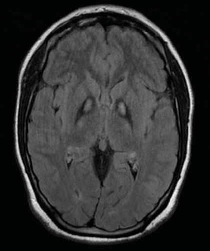
The pharmacological history and the neurological examination suggested a chronic tardive disorder with retrocollis, trunk dystonia, and parkinsonism, which have been frequently described during dopamine receptor antagonist treatments.3 However, this patient required further investigation because of the atypical clinical findings. First, abnormal eye movements were not indicative of antidopaminergic drug use. Second, neuropsychological tests with moderate global cognitive deterioration did not suggest a tardive dystonia. Moreover, the presence of consanguineous parents was consistent with an autosomal recessive disorder. In addition to this, dystonic opisthotonus was recently suggested to be a “red flag” to detect PANK2 mutations in patients with young-onset, complicated dystonia syndromes.3 Therefore, the patient underwent a brain MRI scan that revealed the typical eye-of-the-tiger sign1,2 (Figure 1). Genetic analysis showed that the patient carries a homozygous R286C mutation in the PANK2 gene.

FIGURE 1. T2* MRI Data Show the Eye-of-the-Tiger Sign Produced by a Hypointense Globus Pallidus With a Central Anteromedial Region of Hyperintensity
Conclusions
Although the diverse phenotype of pantothenate kinase-associated neurodegeneration is well known,4 there are few reports investigating possible red flags for neurodegeneration with brain iron accumulation diagnosis, in particular in late-onset (atypical) pantothenate kinase-associated neurodegeneration.1–3 The present clinical observation is in line with the previous suggestion that dystonic opisthotonus can be considered a red flag for neurodegeneration with brain iron accumulation.3 Interestingly, dystonic opisthotonus was the earliest neurological manifestation, whereas other disturbances previously considered typical of pantothenate kinase-associated neurodegeneration (such as oromandibular dystonia) were absent; thus, neurologists must be aware of its differential diagnosis. In addition, eye movements, neuropsychological tests, and family history should always be considered to increase our diagnostic accuracy in neurodegeneration with brain iron accumulation. In conclusion, we aim to stimulate further reports to assess the value of early red flags to detect neurodegeneration with brain iron accumulation.
1 : Genetics and pathophysiology of neurodegeneration with brain iron accumulation (NBIA). Curr Neuropharmacol 2013; 11:59–79Medline, Google Scholar
2 : Syndromes of neurodegeneration with brain iron accumulation (NBIA): an update on clinical presentations, histological and genetic underpinnings, and treatment considerations. Mov Disord 2012; 27:42–53Crossref, Medline, Google Scholar
3 : Dystonic opisthotonus: a “red flag” for neurodegeneration with brain iron accumulation syndromes? Mov Disord 2013; 28:1325–1329Crossref, Medline, Google Scholar
4 : The diverse phenotype and genotype of pantothenate kinase-associated neurodegeneration. Neurology 2005; 64:1810–1812Crossref, Medline, Google Scholar


