Phonagnosia and Inability to Perceive Time Passage in Right Parietal Lobe Epilepsy
To the Editor: Phonagnosia, selective impairment of voice recognition, is a rare, high-order impairment. Apart from a developmental flaw, it can be found in acquired damage of the temporal or parietal lobe, mainly in the right hemisphere.1 The perception of time passing relies mainly on mechanisms of sustained attention.2
Case Report
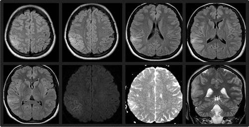
A 25-year-old otherwise healthy epileptic woman was admitted to the hospital after four seizures without recovery of consciousness. She had experienced two previous identical seizures at ages 12 and 14 years. They all began with left oculocephalic deviation and slow left arm rising, followed by generalized tonic-clonic movements. Results on brain CT scan and EEG were normal, and she was treated for 2 years with valproate and was seizure free until age 23 years. At that time, similar seizures restarted, and after normal results on MRI scan and EEG, levetiracetam was started (1000 g/day) with seizure remission. When seen at our unit, the patient underwent a second brain MRI scan, which showed a right cortical parietal T2-hyperintense lesion without gadolinium enhancement and with discrete impairment of water molecule diffusion (Figure 1). An EEG disclosed right frontoparietal slow activity. Phenytoin (in the acute management) and carbamazepine were used, and the patient had complete seizure remission.

FIGURE 1. Brain MRI Scan Showing a Right Cortical Parietal T2-Hyperintense, Nongadolinium-Enhancing Lesion, With Water Molecule Diffusion Restriction
During the following 3 weeks, she started complaining that she could not recognize familiar voices, including those from relatives and famous persons, although she could identify the talking subjects if permitted to see them. She also said that she was incapable of fully acknowledging the passage of time. She was sometimes scared by stimuli that seemed to occur instantaneously but were really several seconds apart. A brain MRI scan 2 months later showed marked improvement, but a residual cortical parietal thickening remained. These data supported the clinical impression of a cortical change secondary to abundant epileptic activity.
Discussion
Phonagnosia was first proposed to encompass disorders of familiar voice recognition, which were found to be more common in right hemispheric lesions, usually involving the parietal lobe.3 Elective impairment in recognizing voices can arise from abnormal processing of complex vocal properties (timbre, articulation, and prosody—elements that distinguish an individual voice).1 Phonagnosia can be a relevant clinical issue, especially in those situations where compensatory cues are unavailable.1
Our patient, whom we believe to have had a postictal parietal cortical dysfunction, was unable to recognize familiar voices, despite normal face recognition as well as environmental and object sound recognition. In a form of associative auditory agnosia, she could not connect the voice with the other semantic-specific information.
In addition, the simultaneous inability to perceive time passage suggests that the same affected parietal circuits are fundamental to this. Indeed, time perception implies encoding time intervals, comparing intervals, and implementing responses, which demand the normal functioning of a broad anatomic network (basal ganglia, right parietal cortex, bilateral premotor cortex, and right dorsolateral prefrontal cortex).2 The disorder presented in our case is distinct from previously reported chronotaraxis, associated with thalamic lesions, and is defined as an isolated disorientation of time.4 Otherwise, as time passage does not have independent sensorial access, it cannot be classified as a true agnosia; therefore, chronoblindness seems a better term to designate this extraordinary inability.
1 : Progressive associative phonagnosia: a neuropsychological analysis. Neuropsychologia 2010; 48:1104–1114Crossref, Medline, Google Scholar
2 : The evolution of brain activation during temporal processing. Nat Neurosci 2001; 4:317–323Crossref, Medline, Google Scholar
3 : Developmental phonagnosia: a selective deficit of vocal identity recognition. Neuropsychologia 2009; 47:123–131Crossref, Medline, Google Scholar
4 : Thalamic chronotaraxis: isolated time disorientation. J Neurol Neurosurg Psychiatry 2007; 78:880–882Crossref, Medline, Google Scholar


