A Magnetization Transfer Analysis of the Thalamus in Schizophrenia
Abstract
The authors investigated the thalamus in schizophrenia by using magnetization transfer ratio (MTR), a novel structural magnetic resonance technique sensitive to subtle neuropathological abnormalities. The dorsomedial nucleus (DMN) and pulvinar were selected because of their connections to limbic, prefrontal, and temporal regions, putatively relevant in schizophrenia. Volume (intracranial; thalamic) and MTR (whole thalamus; DMN; pulvinar) were determined in 25 patients with chronic schizophrenia by DSM-IV criteria and 25 control subjects. There were no significant differences between patients and control subjects in thalamic volume (corrected for intracranial volume) or MTR in whole thalamus, DMN, or pulvinar. No volumetric or MTR abnormalities could be detected in the thalamus of patients with schizophrenia. The findings suggest that abnormalities, if present, are very subtle and beyond the power of resolution of these techniques.
Abnormalities within the thalamus and cortical-subcortical-thalamic circuits serving attention, sensory gating, and information processing have been suggested as a unitary explanation for the symptoms of schizophrenia.1 However, both neuropathological and structural imaging studies have yielded heterogeneous results. Differences in methodology and possibly variability in patient characteristics contribute to the heterogeneous results and limit comparisons between studies.
Neuropathological studies examining the thalamus in schizophrenia are sparse, often involving small elderly populations with a long duration of illness who have received treatments (e.g., electroconvulsive and insulin therapy) with uncertain neuropathological consequences. In addition, these populations often have comorbid illnesses, and studies are further complicated by postmortem variables such as shrinkage. In studies using quantitative methodology, neuronal loss has been reported localized to the pulvinar.2 Stereological studies have reported significant decreases in both dorsomedial nucleus (DMN) volume and DMN cell number, with preserved neuronal density.3–5 Differential neuron loss in the parvocellular and densocellular subnuclei of the DMN has been reported.6 However, others have reported normal total7,8 and thalamic subnuclear8 volumes. Decreased thalamic synaptic density9–11 and decreased neuronal density in the anteroventral nuclei12 have also been reported.
Structural imaging studies have also yielded inconsistent results, reporting both reduced13 and normal14 thalamic volume. Decreased pixel intensity in the lateral thalamus and adjacent white matter upon image averaging1 was not replicated in a more recent study.15 Shape analysis has suggested focal left anterior and right posterior thalamus changes.16,17 Advanced structural magnetic resonance imaging (MRI), with slice resolution now approaching 1 mm, is likely to detect subtle atrophic changes that may have previously been unnoticed. However, subtle neuropathological changes not resulting in atrophy will remain undetected even with this advanced form of conventional MRI.
In vivo magnetic resonance spectroscopy (MRS) has also been used to study thalamic abnormalities in schizophrenia reporting both the presence18 and absence19,20 of reductions in N-acetylaspartate, a marker that is thought to reflect neuronal loss and/or dysfunction. Thalamic hypometabolism has also been reported in functional imaging studies.16,17
Magnetization transfer imaging (MTI) is a novel structural MRI technique capable of detecting subtle neuropathological changes in vivo before atrophy becomes manifest and thus is of particular interest in the study of schizophrenia, where neuropathological changes tend to be subtle.
MTI in the brain is based on the interaction of protons bound to macromolecular structures (e.g., myelin and cell membranes), characterized by restricted motion, and free protons in tissue water, with unrestricted motion. Off-resonance irradiation is applied saturating the magnetization of bound protons, which are not detected by conventional MRI because of their very short relaxation times. There is an exchange of magnetization with free protons, resulting in a decrease in signal intensity that is dependent on the density of macromolecules in the brain. The exchange of magnetization between bound and free protons is measured by the magnetization transfer ratio (MTR). Reduced MTR reflects a reduced capacity of macromolecules to exchange magnetization with tissue water protons and is an index of the integrity of a given tissue.
MT has been used to study white matter diseases such as multiple sclerosis, detecting abnormalities in normal-appearing white21 and gray22 matter that are undetected by use of conventional MRI. Relatively small MTR decreases in the absence of myelin loss23 are likely to reflect decreases in cell membrane protein secondary to neuronal loss, whereas very large decreases reflect myelin damage.24 Neuropathological correlates of MTR in gray matter are not yet fully determined. In healthy volunteers, MTR is highly reproducible,25 is higher in white than in gray matter (because of myelination), and decreases with age.26 In our group's previous studies of chronic schizophrenic patients, we have shown the sensitivity of this technique to detect widespread cortical MTR reductions27 and localized white matter pathology in the temporal lobes.28 In the study described here, we measured MTR in the whole thalamus and two thalamic nuclei (DMN and pulvinar). We used a region of interest (ROI) methodology to identify subtle changes in these two nuclei. The selected nuclei have prefrontal, limbic, and temporal connections. These are considered to be important brain regions in schizophrenia29 and are of sufficient size to accommodate the ROI, standardized at 35.2 mm2. The DMN has connections with prefrontal cortex and amygdala, and the pulvinar has reciprocal connections with auditory association cortex.30 Compared with other gray matter structures, the thalamus has a high MTR, reflecting the presence of myelinated fibers.31 Decreased MTR in these ROIs is likely to reflect neuropathological changes in thalamic myelinated fibers and/or thalamic neuronal atrophy or loss. The null hypothesis is that there will be no differences in MTR between patients and control subjects.
METHODS
Subjects
Twenty-five patients (19 males, 6 females), mean age 37.2 years (range 25–46), who fulfilled DSM-IV criteria for schizophrenia were recruited from the Bethlem and Maudsley Hospitals. Patients were assessed by an experienced research psychiatrist (J.F.) using a clinical interview and careful review of patient records. Most patients (n=23) were of the paranoid subtype. Mean symptom duration was 14.3 years (range 3–22). All patients were receiving neuroleptic medication. Five patients were being treated with atypical medication, but all had previously been treated with typical neuroleptics. Mean dosage (chlorpromazine equivalents; British National Formulary, 2000) was 367.5 mg/day (range 37.5–700 mg/day). No patients had been exposed to electroconvulsive treatment. Twenty-five healthy control subjects (19 males, 6 females), mean age 35.2 years (range 24–49), were also selected. Subjects with a history of neurological or systemic illness, head injury, drug abuse, or alcohol dependence were excluded. Schizophrenic patients and control subjects were matched for age, gender, and parental social class.32 The study received approval from the ethics committees of the Bethlem and Maudsley NHS Trust, Institute of Neurology, and National Hospital for Neurology and Neurosurgery, London. Written consent was obtained from all subjects. These subjects had participated in our earlier studies.27,28
Handedness was assessed by using the Annett Questionnaire.33 Most subjects were right-handed (schizophrenic, n=24; control, n=24). Premorbid IQ was assessed by using the National Adult Reading Test (NART)34 and was available for 19 patients (mean score 110.7) and 19 control subjects (mean score 113.2). Positive and negative subscales of the Positive and Negative Syndrome Scale (PANSS)35 were used to assess symptom severity during the week prior to scanning in schizophrenic patients. Most patients scored higher on the negative than the positive subscale of the PANSS, reflecting a preponderance of negative symptoms (mean total scores of 18.6 and 11.6, respectively).
Imaging
A GE Signa 1.5-tesla MR scanner with standard quadrature head coil was used. T2-weighted and proton density images were acquired, using a dual echo sequence (echo time [TE]=15/90 ms, repetition time [TR]=3,000 ms, 28 contiguous 5-mm axial slices, 256×256 pixel image matrix, 24×24 cm2 field of view). A spin-echo sequence (TE=30/80ms, TR=1,720 ms, 28 contiguous 5-mm axial slices, 256×128 pixel image matrix, 24×24 cm2 field of view) both with and without an MT saturation pulse (16 ms, 23.2 microtesla Hamming appodized 3-lobe sinc pulse, applied 1 kHz from water resonance) was used for MT. The MT images were co-registered with the T2-weighted and proton density images.36,37 MTR was calculated on a pixel-by-pixel basis from the formula MTR=[Mo−Ms] / [Mo]×100, where Ms and Mo are the mean signal intensities with and without the saturation pulse, respectively.
Image Analysis
Data were processed on a Sun Ultra workstation (Sun Microsystems, Mountain View, CA), using the DispImage software package.38 Intracranial volume included cerebrum, cerebellum, ventricles, and cerebrospinal fluid within the sulci and was determined by using a semi-automated technique. The cumulative total area of each 5-mm axial slice was multiplied by slice thickness to determine volume.
Thalamic boundaries and ROI localization (Figure 1) were determined by using protocols developed in conjunction with a neuroradiologist (G.d.B.) with reference to standard atlases.39,40 The thalamic boundaries were as follows: anterolateral: head of caudate; anteromedial: third ventricle; lateral: internal capsule; posterior: choroidal fissure of the lateral ventricle; medial: third ventricle; superior: body of the lateral ventricle; inferior: last slice in which thalamus was visible superior to the first slice in which both red nucleus and substantia nigra were visible.
Thalamic volume was determined by manually tracing thalamic margins on all slices in which the thalamus was visible (usually 4 to 5 slices) and multiplying cumulative total area by slice thickness. Mean thalamic MTR was determined by averaging cumulative MTR values for all thalamic slices.
Thalamic subnuclear margins are difficult to visualize with MRI. Thus, relatively large, anatomically relevant subnuclei were selected that could accommodate the ROI. Bilateral rectangular 35.2-mm2 ROIs were manually placed in 2 consecutive slices containing the largest thalamic area (immediately superior to the anterior commissure) strictly according to the protocol (Figure 1) by a rater (M.S.B.) who was blind to group membership. The ROIs were placed on the T2-weighted image to avoid bias in placement, after adjacent slices were examined to minimize cerebrospinal fluid partial volume effects. The MT images were co-registered with T2-weighted images,36,37 allowing ROIs to be automatically transferred onto the MT images. The accuracy of ROI placement was qualitatively assessed by a neuroradiologist (G.d.B.) on random sampling of 6 controls and 6 patients. Mean MTR was determined for DMN and pulvinar by averaging MTR in both slices for each nucleus.
Interrater reliability for MTR ROIs was assessed for MTR values obtained in a random sample of 6 control subjects and 6 patients by 2 raters (M.S.B. and J.F.). Intraclass correlation coefficients ranged from 0.90 to 0.93 for all ROIs.
RESULTS
There were no significant differences in age, gender, handedness, or parental social class between patients and control subjects.
Volume Analysis
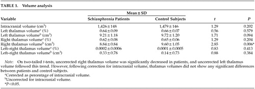
Two-tailed t-tests revealed no significant differences for intracranial volume or thalamic volume corrected for intracranial volume (Table 1). Schizophrenic brains were 3.5% smaller on the basis of mean values, but this was not significant (P=0.202). Right thalamus volume was reduced in patients (P=0.006) on the basis of uncorrected volume analysis, and a similar trend was seen for left thalamus (P=0.094), but this reduction was not preserved after correction for intracranial volume.
MTR Analysis
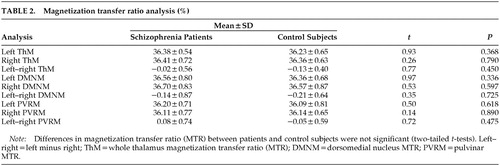
Two-tailed t-tests did not reveal significant MTR differences between the two groups for whole thalamus, DMN, or pulvinar. Left minus right MTR values for the DMN and pulvinar were also similar, suggesting there were no differences in hemispheric asymmetry in the two groups (Table 2). Using Spearman's correlation coefficient, we failed to find significant correlations of NART-estimated premorbid IQ or PANNS subscales of positive and negative symptoms with MTR in the pulvinar or DMN.
DISCUSSION
Using a novel MRI technique sensitive to the presence of subtle neuropathological abnormalities, we failed to detect thalamic volumetric or MTR reductions in patients with chronic schizophrenia compared with matched control subjects. In this study we focused on two thalamic nuclei with frontolimbic (DMN) and temporal lobe (pulvinar) connections putatively relevant in schizophrenia. The normal MTR values in the thalamus as a whole suggest that significant abnormalities in other individual thalamic nuclei not explored in this study are unlikely. Within-group comparisons of gender effects (6 females) or schizophrenia subtype were not possible because of the small sample size and the fact that most patients were of the paranoid subtype. Therefore it is not possible to say whether MTR abnormalities may have been present in other clinical subgroups. We also failed to find correlations between NART-estimated premorbid IQ or PANNS subscales for positive and negative symptoms and MTR in the pulvinar or DMN. However, because of our small sample size we cannot exclude the possibility of thalamic abnormalities in subgroups of patients with earlier-onset schizophrenia, as suggested by some studies,41 or in those with specific symptom clusters or cognitive impairment.
The neuropathological correlates of MTR are not fully understood, but there is substantial evidence that MTR is sensitive to changes in tissue organization and macromolecular structure.21–23 Medial temporal cortical27 and temporal lobe white matter28 MTR abnormalities have been demonstrated in the same patients, supporting our view that the technique is sufficiently sensitive to detect the subtle neuropathological abnormalities in schizophrenia.
Differences in thalamic volumes between our patients and control subjects did not survive correction for intracranial volume. Better slice resolution may have increased our chances of detecting volumetric differences between the two groups, although a high-resolution study using 1.5-mm contiguous slices in a sample of 15 right-handed patients with chronic schizophrenia also reported no differences in thalamic volumes between the patients and control subjects.14 Others using lower-resolution image acquisition have reported significant thalamic volume reductions in schizophrenic patients,13 and these conflicting results may, in part, be attributed to methodological differences including border definition, signal-to-noise ratio, and slice acquisition parameters. The use of high-resolution techniques (i.e., CAIR, or cortex-attenuated inversion recovery MRI sequence) that allow visualization of specific thalamic nuclei42 may help to settle the differences between various studies.
The main limitations of our study are the small sample size, possible partial volume effects, and limited resolution dictated by 5-mm contiguous slices that may not be sensitive to subtle localized volume changes. A previous study by our group28 reported a 2% difference in MTR between chronic schizophrenic patients and control subjects in the right temporal lobe, which was highly significant, and a retrospective analysis revealed that 16 patients and 16 control subjects would have been sufficient to detect differences with a power of 80% and a significance level of 0.05. In the present study, the maximum difference between patients and control subjects for MTR was in the left DMN (0.20%), and the standard deviation for control subjects was 0.68%. On the basis of these variables, if significant differences were present between patients and control subjects in the left DMN, we would require a sample of 400 subjects to detect a difference with a power of 80% and a significance level of 0.05. Larger samples would be required for the other nuclei tested in this study. Arguably, differences of this magnitude may not be biologically relevant.
A better understanding of the neuropathological correlates of MTR is required before our findings can be interpreted conclusively. However, with these caveats and the need for replication in mind, our results do not support the presence of gross pathological changes in the thalamus. If present, thalamic abnormalities are likely to be far more subtle than those present in the cortex in chronic schizophrenia that are detectable with the same technique. Our findings do not exclude state-dependent abnormalities in functional connectivity43 but suggest that these are more likely to result from cortical than thalamic abnormalities.
ACKNOWLEDGMENTS
The authors are grateful to members of the NMR Unit for their assistance and also thank the patients and control subjects who participated. This study was supported by a grant from the Wellcome Trust.

FIGURE 1. Region of interest (ROI) localization in dorsomedial nucleus (DMN) and pulvinar. In the pulvinar, ROIs were placed equidistant from the third ventricle (medially) and internal capsule (laterally), immediately anterior to the choroidal fissure of the lateral ventricle. In the DMN, ROIs were equidistant from the head of caudate (anterolaterally); the margin of the third ventricle (anteromedially); and the choroidal fissure of the lateral ventricle (posteriorly), immediately lateral to the third ventricle.
 |
 |
1 Andreasen NC, Arndt S, Swayze V, et al: Thalamic abnormalities in schizophrenia visualized through MRI averaging. Science 1994; 266:294-298Crossref, Medline, Google Scholar
2 Dom R, de Saedeler J, Bogerts B, et al: Quantitative cytometric analysis of basal ganglia in catatonic schizophrenics, in Biological Psychiatry, edited by Perris C, Struwe G, Jansson B. Amsterdam, Elsevier, 1981, pp 723-726Google Scholar
3 Pakkenberg B: Pronounced reduction of total neuron number in mediodorsal thalamic nucleus and nucleus accumbens in schizophrenics. Arch Gen Psychiatry 1990; 47:1023-1028Crossref, Medline, Google Scholar
4 Pakkenberg B: The volume of the mediodorsal thalamic nucleus in treated and untreated schizophrenics. Schizophr Res 1992; 7:95-100Crossref, Medline, Google Scholar
5 Young K, Manaye K, Liang CL, et al: Reduced number of mediodorsal and anterior thalamic neurons in schizophrenia. Biol Psychiatry 2000; 47:944-953Crossref, Medline, Google Scholar
6 Popken G, Bunney W, Potkin S, et al: Subnucleus-specific loss of neurons in medial thalamus of schizophrenics. Proc Natl Acad Sci USA 2000; 97:9276-9280Crossref, Medline, Google Scholar
7 Rosenthall R, Bigelow LB: Quantitative brain measurements in chronic schizophrenia. Br J Psychiatry 1972; 121:259-264Crossref, Medline, Google Scholar
8 Lesch A, Bogerts B: The diencephalon in schizophrenia: evidence for reduced thickness of the periventricular grey matter. Eur Arch Psychiatr Neurol Sci 1984; 234:212-219Crossref, Medline, Google Scholar
9 Blennow K, Davidson P, Gottfries C-G, et al: Synaptic degeneration in the thalamus in schizophrenia. Lancet 1996; 348:692-693Crossref, Medline, Google Scholar
10 Blennow K, Bogdanovic N, Helig M, et al: Reduction of the synaptic protein Rab3a in the thalamus and connecting brain regions in post-mortem schizophrenic brains. J Neural Transm 2000; 107:1085-1097Crossref, Medline, Google Scholar
11 Davidsson P, Gottfries J, Bogdanovic N, et al: The synaptic-vesicle-protein proteins Rab3a and synaptophysin are reduced in thalamus and related cortical brain regions in schizophrenic brains. Schizophr Res 1999; 40:23-29Crossref, Medline, Google Scholar
12 Danos P, Bruno B, Bernstein H-G, et al: Schizophrenia and anteroventral thalamic nucleus: selective decrease of parvalbumin-immunoreactive thalamocortical projection neurons. Psychiatry Res 1998; 82:1-10Crossref, Medline, Google Scholar
13 Flaum MD, Swayze VW, O'Leary DS, et al: Effects of diagnosis, laterality and gender on brain morphology in schizophrenia. Am J Psychiatry 1995; 152:704-714Crossref, Medline, Google Scholar
14 Portas CM, Goldstein JM, Shenton ME, et al: Volumetric evaluation of the thalamus in schizophrenic male patients using MRI. Biol Psychiatry 1998; 43:649-659Crossref, Medline, Google Scholar
15 Wolkin A, Rusinek H, Vaid G, et al: Structural magnetic resonance image averaging. Am J Psychiatry 1998; 155:1064-1073Crossref, Medline, Google Scholar
16 Buschbaum MS, Someya T, Teng CY, et al: PET and MRI of the thalamus in never-medicated patients with schizophrenia. Am J Psychiatry 1996; 153:191-199Crossref, Medline, Google Scholar
17 Hazlett EA, Buschbaum MS, Byne W, et al: Three-dimensional analysis with MRI and PET of the size, shape and function of the thalamus in the schizophrenia spectrum. Am J Psychiatry 1999; 156:1190-1199Medline, Google Scholar
18 Diecken R, Johnson C, Eliaz Y, et al: Reduced concentrations of thalamic N-acetylaspartate in male patients with schizophrenia. Am J Psychiatry 2000; 157:644-647Crossref, Medline, Google Scholar
19 Bertolini A, Callicott JH, Elman I, et al: Regionally specific neuronal pathology in untreated patients with schizophrenia: a proton MRS imaging study. Biol Psychiatry 1998; 43:641-648Crossref, Medline, Google Scholar
20 Bertolini A, Nawroz S, Mattay VS, et al: Regionally specific pattern of neurochemical pathology in schizophrenia as assessed by multislice proton MRS imaging. Am J Psychiatry 1996; 153:1554-1563Crossref, Medline, Google Scholar
21 Filippi M, Campi A, Dousset V: A magnetization transfer imaging study of normal-appearing white matter in multiple sclerosis. Neurology 1995; 45:478-482Crossref, Medline, Google Scholar
22 Cercignani M, Bozzali M, Iannucci G, et al: Magnetisation transfer ratio and mean diffusivity of normal appearing white and grey matter from patients with multiple sclerosis. J Neurol Neurosurg Psychiatry 2001; 70:311-317Crossref, Medline, Google Scholar
23 Brochet B, Dousset V: Pathological correlates of magnetization transfer imaging abnormalities in animal models and humans with multiple sclerosis. Neurology 1999; 53(suppl):S12-S17Google Scholar
24 Van Waaesberghe JH, Kamphorst W, Van Waldervwen MA: Histopathological correlates of MTR and hypointense signal intensity on T1 SE in multiple sclerosis lesions: a direct postmortem study (abstract). Multiple Sclerosis 1998; 4:272Google Scholar
25 Barker GJ, Tofts PS, Gass A: An interleaved sequence for accurate and reproducible clinical measurement of magnetization transfer ratio. Magn Reson Imaging 1996; 14:403-411Crossref, Medline, Google Scholar
26 Silver NC, Barker GJ, MacManus DG, et al: Magnetisation transfer ratio of normal brain white matter: a normative database spanning four decades of life. J Neurol Neurosurg Psychiatry 1997; 62:22-38Crossref, Medline, Google Scholar
27 Foong J, Symms MR, Barker GJ, et al: Investigating neuropathological abnormalities in schizophrenia using magnetisation transfer imaging (abstract). Proceedings of the International Society of Magnetic Resonance Medicine 1999; 7:949Google Scholar
28 Foong J, Maier M, Barker G, et al: In vivo investigation of white matter pathology in schizophrenia with magnetisation transfer imaging. J Neurol Neurosurg Psychiatry 2000; 68:70-74Crossref, Medline, Google Scholar
29 Wright IC, Ellison ZR, Sharma T, et al: Mapping of grey matter changes in schizophrenia. Schizophr Res 1999; 35:1-14Crossref, Medline, Google Scholar
30 Crosson B, Hughes CW: Role of the thalamus in language: is it related to schizophrenic thought disorder? Schizophr Bull 1987; 13:605-621Crossref, Medline, Google Scholar
31 Mehta RC, Pike B, Enzmann DR: Magnetization transfer MR of the normal adult brain. Am J Neuroradiol 1995; 16:2085-2091Medline, Google Scholar
32 Goldthorpe JH, Hope K: The Social Grading of Occupations: A New Approach and Scale. Oxford, UK, Clarendon Press, 1974Google Scholar
33 Annett MA: A classification of hand preference by association analysis. Br J Psychol 1970; 61:303-321Crossref, Medline, Google Scholar
34 Nelson H, Willison J: The National Adult Reading Test (NART), 2nd edition. Windsor, UK, NFER-Nelson, 1991Google Scholar
35 Kay SR, Fiszbein A, Opier LA: The Positive and Negative Syndrome Scale (PANSS) for schizophrenia. Schizophr Bull 1987; 13:261-276Crossref, Medline, Google Scholar
36 Woods R, Cherry S, Mazziotta J: Rapid automated algorithm for aligning and reslicing PET images. J Comput Assist Tomogr 1992; 16:620-633Crossref, Medline, Google Scholar
37 Symms M, Barker G, Holmes A: A rapid automated system for detection of serial changes in transient ischaemic attack using registration and subtraction of three dimensional images (abstract). Proc Intl Soc Mag Reson Med 1997, no 584Google Scholar
38 Plummer DL: DispImage: a display and analysis tool for medical images. Rev Neuroradiol 1992; 5:489-495Google Scholar
39 Stelmasiak M: Anatomical Atlas of the Human Brain and Spinal Cord. Warsaw, Polish State Medical Publishers, 1956Google Scholar
40 Hanaway J, Woolsey TA, Mokhtar HG, et al: The Brain Atlas. Bethesda, MD, Fitzgerald Scientific Press, 1998Google Scholar
41 Frazier JA, Giedd JN, Hamburger SD, et al: Brain anatomic magnetic resonance imaging in childhood-onset schizophrenia. Arch Gen Psychiatry 1996; 53:617-624Crossref, Medline, Google Scholar
42 Magnotta V, Gold S, Andreasen N, et al: Visualization of subthalamic nuclei with cortex attenuated inversion recovery MR imaging. Neuroimage 2000; 11:341-346Crossref, Medline, Google Scholar
43 Spence SA, Liddle MD, Stefan MD, et al: Functional anatomy of verbal fluency in people with schizophrenia and those at genetic risk: focal dysfunction and distributed disconnectivity reappraised: a PET study of verbal fluency. Br J Psychiatry 2000; 176:52-60Crossref, Medline, Google Scholar