Frontal Lobe Dysfunction in Panic Disorder: a Comparison of Multichannnel Near-infrared Spectroscopy in Monozygotic Twins Discordant for Panic Disorder
To the Editor: Recent studies speculate that frontal lobe dysfunction in panic disorder plays a certain role to modulate anxiety responses. 1 This is the first case report about the use of near-infrared spectroscopy to investigate frontal lobe function in monozygotic twins discordant for panic disorder.
Case Report
The subjects were 30-year-old male monozygotic twins discordant for panic disorder as diagnosed by the DSM-IV. The affected twin (Twin A) was diagnosed with panic disorder without agoraphobia at age 25. The unaffected subject (Twin B) was healthy and works as a teacher of a high school and revealed no current or past psychiatric problems. The study participants included 24 right-handed male patients with panic disorder with or without agoraphobia (mean age ± SD=31.46±4.78, mean score of the Panic Disorder Severity Scale =10.21±5.80) diagnosed according to the criteria in the DSM-IV and 20 right-handed healthy male volunteers (mean age=29.95±5.44) without psychiatric disorder as the comparison group. The changes in the concentration of oxygenated hemoglobin in the panic disorder group were significantly smaller than in the healthy comparison group.
The word fluency task used here consisted of a 30-second pretask baseline period, a 60-second task period consisting of three 20-second blocks, and a 70-second posttask baseline period. Relative oxygenated hemoglobin concentration changes were measured with a 52-channel near-infrared spectroscopy machine (Hitachi ETG-4000). Of 52 channels, the mean changes in oxygenated hemoglobin by three regions were calculated: 10 right lateral prefrontal anterior (PFC), 14 anterior PFC, and 10 left lateral PFC channels.
The numbers of words generated during the word fluency task were nine words in Twin A and nearly the same number of words in Twin B (10 words). The twins generated words less than average on the comparison sample (mean words= 13.37±4.15). The oxygenated hemoglobin changes were small in the anterior PFC in Twin A as much as Twin B. It was approximately the same activation in the panic disorder group ( Figure 1 , panel B). On the other hand, in the bilateral PFC, the [oxy-Hb] changes were small in Twin A while the oxygenated hemoglobin changes were distributed at the midpoint of the comparison groups in Twin B ( Figure 1 , panels A and C).
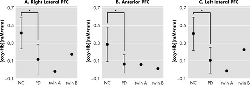
Vertical bars represent the standard deviation.
*p<0.001 by t-test, respectively
NC=normal comparison subject; PD=panic disorder; twin A=affected by panic disorder; twin B=unaffected by panic disorder
Discussion
Our presented results provide the possibility that the oxygenated hemoglobin changes in the frontal lobe reflect a pathophysiological feature of panic disorder. Previous research in a study of twins showed that the frontal cortex was markedly influenced by genetic factors. 2 In this present case, the findings suggest that the smaller activation in the anterior prefrontal cortex could possibly be associated with a genetic factor as an endophenotype for panic disorder. In contrast, the differences in the bilateral prefrontal cortex might correspond to a state marker. Further research of frontal lobe function will help elucidate the pathophysiology of panic disorder.
1. Gorman JM, Kent JM, Sullivan GM, et al: Neuroanatomical hypothesis of panic disorder, revised. Am J Psychiatry 2000; 157:493–505Google Scholar
2. Thompson PM, Cannon TD, Narr KL, et al: Genetic influences on brain structure. Nat Neurosci 2001; 4:1253–1258Google Scholar