Cerebral Amyloid Angiopathy-Related Inflammation: A Treatable Cause of Rapidly-Progressive Dementia
Case Report
An 83-year-old woman presented with rapidly-progressive dementia, global aphasia, and cerebellar ataxia. Blood pressure, standard laboratory, and CSF examination were normal. The constellation of rapid cognitive decline and imaging findings of extensive asymmetrical subcortical areas of T2-hyperintensity and multiple cortico-subcortical microbleeds was considered sufficient to reach a presumptive diagnosis of cerebral amyloid angiopathy (CAA)-related inflammation.1,2 A course of IV high-dose steroid treatment, followed by prolonged oral tapering, led to a dramatic clinical improvement and complete resolution of the vasogenic edema on MRI. Given the noticeable response to treatment, a diagnostic brain biopsy had not subsequently been carried out. During a follow-up period of 18 months, the patient remained stable without maintenance treatment.
Discussion
Cerebral amyloid angiopathy (CAA) is characterized by deposition of amyloid protein in vessel walls of leptomeningeal and cortical small arteries; it was first described in 1907.1 A variety of distinct clinical phenotypes have been attributed to this CNS vasculopathy, with spontaneous recurrent lobar hemorrhage in the elderly patient being the most widely recognized.1 More recent studies have established that, in CAA associated with leukoencephalopathy, an inflammatory variant of CAA can be distinguished.2–4 Mean age at disease onset is about 67 years.1–4 A clinico-radiological-histopathological syndrome has been described, consisting of a clinical phenotype of rapidly-progressive dementia, extensive and markedly asymmetric T2 hyperintense white-matter lesions, together with multiple scattered cortical/subcortical microbleeds on MR-imaging (Figure 1) and typical vascular amyloid deposits associated with perivascular inflammatory changes on histopathological examination.2–4 Although the diagnostic gold standard remains histopathological examination, it has been suggested that in the appropriate clinical setting, typical imaging findings, as outlined above, might be sufficient to diagnose CAA-related inflammation, without necessitating brain biopsy.3,5 A diagnostic entity termed “probable CAA-related inflammation” has been proposed.3,5 Thus, initiating a short treatment trial with high-dose steroids, without performing brain biopsy, can be done if further studies (e.g., blood pressure, laboratory, CSF) make an infectious, hypertensive, or neoplastic etiology unlikely. Most patients respond well to immunosuppressive therapy in the acute phase; only a few need long-term maintenance therapy.2
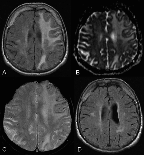
A. T2-FLAIR weighted images at initial evaluation showing extensive subcortical signal prolongation, extending from the ventricular margins to the subcortical u-fibers, with only minor mass effect. Contrast enhancement was not observed (not shown).
B. The apparent diffusion-coefficient map, derived from diffusion-weighted imaging, correspondingly shows increased diffusivity consistent with vasogenic edema.
C. T2*-weighted gradient-echo images demonstrate multiple scattered cortical and subcortical microbleeds. No microbleeds could be detected in basal ganglia, thalamus, or cerebellum (not shown).
D. T2-weighted scan performed after treatment with high-dose steroids shows complete resolution of the vasogenic edema. Remaining areas of focal T2 hyperintensity may either represent residual gliosis or unrelated microangiopathic or age-associated changes.
The first priority of diagnostic evaluation of dementia syndrome is the identification of potentially treatable causes. Cerebral amyloid angiopathy (CAA)-related inflammation is associated with a clinical phenotype of rapidly-progressive dementia, and most patients respond well to immunosuppressive therapy. It has been suggested that it might be diagnosed non-invasively, based on a constellation of typical clinical and radiological findings. Thus, careful neuroradiological evaluation for microbleeds in patients with rapidly-progressive dementia and leukoencephalopathy can help to define a potentially treatable cause of rapidly-progressive dementia syndrome.
1. : Cerebral amyloid angiopathy and lobar intracerebral hemorrhage. Arch Neurol 2006; 63:148–151Crossref, Medline, Google Scholar
2. : Clinical manifestations of cerebral amyloid angiopathy-related inflammation. Ann Neurol 2004; 55:250–256Crossref, Medline, Google Scholar
3. : Course of cerebral amyloid angiopathy-related inflammation. Neurology 2007; 68:1411–1416Crossref, Medline, Google Scholar
4. : Aβ-related angiitis: primary angiitis of the central nervous system associated with cerebral amyloid angiopathy. Brain 2005; 128:500–515Crossref, Medline, Google Scholar
5. : Cerebral amyloid angiopathy-related inflammation: three case reports and a review. J Neurol Neurosurg Psychiatry 2011; 82:20–26Crossref, Medline, Google Scholar