A Case of Schizophrenia Accompanied By Lissencephaly
To The Editor: Lissencephaly is one of the cortical dysplasia diseases that include schisencephaly, polymicrogria, and heterotopic gray matter. These diseases are caused by defective neuronal migration. Cortical dysplasia is often accompanied by severe mental retardation and intractable epilepsy; however, there are very few reported cases of cortical dysplasia with psychotic symptoms,1,2 and, before the present case, we found no reports on lissencephaly with psychotic symptoms. Here, we experienced a case of schizophrenia accompanied by lissencephaly with cerebellar hypoplasia.
Case report
A 32-year-old woman presented with auditory hallucinations and persecutory delusions. She had been suffering from hyperacusis since high school and showed spiritual inclinations, although she had not experienced psychotic symptoms until the age of 32 years. She had no family history of psychiatric disorders, developmental problems, or epilepsy.
The patient was clinically diagnosed with schizophrenia by a psychiatrist 2 weeks before hospitalization, without any diagnostic brain imaging, and she was prescribed an antipsychotic (aripiprazole); however, it did not improve her symptoms. She was admitted to our hospital, and we also diagnosed her with paranoid-type schizophrenia, according to the Diagnostic and Statistical Manual–IV, and we prescribed risperidone, which was increased to 6 mg. After 4 weeks, we changed risperidone to olanzapine and increased the dosage to 15 mg; however, this change was ineffective.
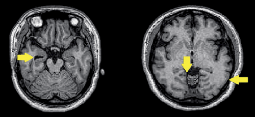
We conducted magnetic resonance imaging (MRI) 5 weeks after hospitalization, because the patient had refused MRI at the time of hospitalization. The MRI showed mild lissencephaly, which was dominant in the temporal and occipital lobes; mild atrophy of the right hippocampus; and hypoplasia of the cerebellum (Figure 1).
Although her electroencephalogram (EEG) did not show convulsive spikes or any other abnormalities except low amplitude, we decided to prescribe sodium valproate on the basis of a similar case report.2 After this prescription, her auditory hallucinations and persecutory delusions improved.
Comment
Typical lissencephaly is divided into agyria–pachygyria spectrum and cobblestone lissencephaly. Lissencephaly can also be accompanied by cerebellar hypoplasia, which is caused by dysfunction of the reelin gene. Although we were unaware whether our patient had this gene dysfunction, this gene is a controlling factor in cortical structure. This gene also has a possible relationship with schizophrenia;3,4 however, no cases of lissencephaly with schizophrenia have been reported. Although we had not found any study or case report about schizophrenia associated with lissencephaly, we found one report about schizophrenia associated with schizencephaly.2 This case report indicated that the patient was resistant to antipsychotics, and that her psychotic symptoms and abnormal EEG improved with carbamazepine administration, although the patient did not have a history of epilepsy.
MRI is becoming common in clinical psychiatry, and it detects many more cases of cortical dysplasia than expected. Thus, we may encounter more cases of cortical dysplasia with psychosis in the future.
1 : Schizencephaly associated with psychosis. J Neurol Neurosurg Psychiatry 1997; 63:373–375Crossref, Medline, Google Scholar
2 : [A cases of schizencephaly associated psychosis]. No To Shinkei 2006; 58:611–614Medline, Google Scholar
3 : Decrease in reelin and glutamic acid decarboxylase67 (GAD67) expression in schizophrenia and bipolar disorder: a postmortem brain study. Arch Gen Psychiatry 2000; 57:1061–1069Crossref, Medline, Google Scholar
4 : Down-regulation of dendritic spine and glutamic acid decarboxylase 67 expressions in the reelin haploinsufficient heterozygous reeler mouse. Proc Natl Acad Sci USA 2001; 98:3477–3482Crossref, Medline, Google Scholar