Neurosyphilis Presenting as Primary Progressive Aphasia
To the Editor: Primary progressive nonfluent aphasia (PPnfA) is an aphasic disorder that is classified in the spectrum of frontotemporal dementia diseases.1 It is characterized by disturbed grammatical comprehension and expression, as well as reduction of speech sound production. We present a adult man whose initial symptoms and signs were compatible with PPnfA in whom later work-up surprisingly unmasked neurosyphilis.
Case Report
A 52-year-old normotensive native man was admitted from the outpatient memory clinic of our department for evaluation of speech difficulties that started 6 months earlier. His past medical history was otherwise unremarkable. Neurological examination revealed mild cogwheel rigidity, positive grasp reflexes, and bilateral Babinski signs. MMSE score was 28, and the Addenbrooke Cognitive Examination–Revised (ACE–R) test score was 74/100. He presented reduced verbal fluency, with word-finding difficulties. Brain MRI showed bilateral perisylvian cortical atrophy, more prominent in the left hemisphere (Figure 1), and routine EEG revealed theta-wave activity in the left frontotemporal area. Extensive serology and hematological tests were normal except for positive Venereal Disease Research Laboratory (VDRL) test (titer: 1/128). Subsequently, serum tests for rapid plasma regain (RPR) and fluorescent treponemal antibody absorption (FTA-Abs) were both positive, with RPR titer: 1/128. Spinal tap demonstrated clear CSF with 160 cells/mm3 (50% neutrophils), along with protein: 135 mg/dl, glucose: 28 mg/dl, and positive VDRL (1:16), RPR (1:32), and positive FTA-Abs.
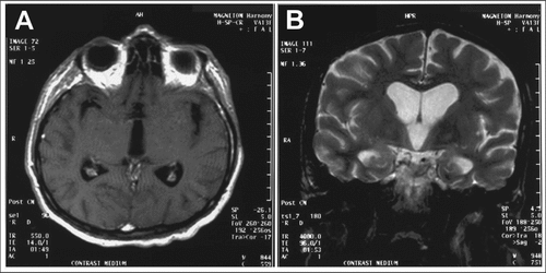
[A]: T1-weighted image with contrast medium; [B]: T2-weighted image shows ventricular enlargement due to parenchymal atrophy, incompatible with his age.
The patient was treated with IV 4×10⁶ units of penicillin administrated 6 times daily for 15 days and followed by 2.4 million units of benzathine penicillin 5 IM once a week for 3 weeks. A second spinal tap showed clear, acellular CSF, with protein 95 mg/dl, glucose 37 mg/dl, positive VDRL (1:8), and RPR (1:16).
On follow-up 6 months later, a significant improvement of language production was observed; his ACE–R score was 92/100; MMSE score was 30; and the rest of the neurological signs remained unchanged.
Discussion
Neurosyphilis can present with progressive cognitive deterioration accompanied by psychiatric manifestations,2 and it is considered one of the treatable and reversible causes of dementia.3 With the present progress of socioeconomic globalization, the spread of migration from developing countries, and the recently-published increasing spread of syphilis,4 we have to reconsider neurosyphilis even in immunocompetent adults. The correct diagnosis could be crucial because early therapy could dramatically affect prognosis.5 Also, our case was atypical because it presented with progressive aphasic disturbances resembling PPnfA. Had we omitted the special serological tests for syphilis, our case could had probably remained undiagnosed.
In conclusion, syphilis should be included in the differential diagnosis of early-onset dementias, even if the initial symptoms and neuroimaging findings point toward a degenerative dementing process.
1 : Classification of primary progressive aphasia and its variants. Neurology 2011; 76:1006–1014Crossref, Medline, Google Scholar
2 : Neurosyphilis: MRI features and their phenotypic correlation in a cohort of 35 patients from a tertiary care university hospital. Neuroradiology 2013; 55:379–388; available at doi: 10.1007/s00234-012-1017-9Crossref, Medline, Google Scholar
3 : Clinical presentation and imaging of general paresis due to neurosyphilis in patients negative for human immunodeficiency virus. J Clin Neurosci 2010; 17:308–310Crossref, Medline, Google Scholar
4 : Resurgent syphilis in the United States: urgent need to address an evolving epidemic. Ann Intern Med 2011; 155:192–193Crossref, Medline, Google Scholar
5 : Review: Neurosyphilis: a historical perspective and review. CNS Neurosci Ther 2010; 16:e157–e168Crossref, Medline, Google Scholar