The American Psychiatric Association (APA) has updated its Privacy Policy and Terms of Use, including with new information specifically addressed to individuals in the European Economic Area. As described in the Privacy Policy and Terms of Use, this website utilizes cookies, including for the purpose of offering an optimal online experience and services tailored to your preferences.

Please read the entire Privacy Policy and Terms of Use. By closing this message, browsing this website, continuing the navigation, or otherwise continuing to use the APA's websites, you confirm that you understand and accept the terms of the Privacy Policy and Terms of Use, including the utilization of cookies.

×
LettersFull Access

A Case of Visual Hypoemotionality Induced by Interferon Alpha-2b Therapy in a Patient With Chronic Myeloid Leukemia

Published Online:

To the Editor: We describe the case of a 41-year-old woman affected by chronic myeloid leukemia, who underwent a standard treatment with Interferon alpha-2b (IFNα2b) 4 months before our observation, with good clinical and hematological response. After the first dose of therapy, she began to report difficulties in memory and concentration and “emotional hyporeactivity.” The patient had no history of pretreatment neurologic or psychiatric disease, and she denied any similar affliction in her family. Neurological examination was normal. Her “emotional hyporeactivity” consisted in lack of vividness and affective coloring of visual stimuli, with preserved emotional responses to nonvisual stimuli, in accord with a diagnosis of Visual Hypoemotionality (VH). Biochemical and immunological examinations on blood and CSF were unremarkable. Standard EEG, visual acuity, visual field, VEP, cranial CT scan, and cerebral MRI (T1, T2, DWI) did not show any abnormalities. A complete neuropsychological assessment showed a selective deficit of visuospatial memory and a mild prosopagnosia (Rey's Complex Figure Delayed Recall: score 5.5, cutoff: ≥15.93; Recognition of Unknown Faces Test: score 36, cutoff ≥41). A 99m ECD-SPECT scan showed a focal hypoperfusion of the right temporo-mesial region (Figure 1). At 1-year follow-up, a complete remission of symptoms was observed, and both technetium-99m ECD-SPECT scan and neuropsychological evaluation were normal.

FIGURE 1. Technetium-99m ECD-SPECT Scan Showing Hypoperfusion of Right Temporo-Mesial Region

Discussion

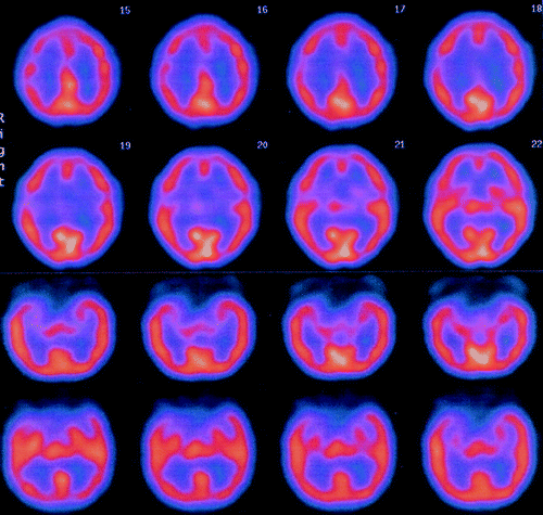
Neuropsychiatric toxicity has been often described in patients receiving IFN-α2b. The most frequently reported adverse effects are moderate anxiety or depression, changes in mood, and difficulties in memory and concentration, which usually disappear after the therapy is discontinued; more severe complications are uncommon, and include frank paranoia, dementia, coma, seizures, and neuropathy. The mechanisms underlying these effects are still not clear.1 Our patient presented with so-called “Visual Hypoemotionality,” a rare syndrome characterized by a disruption of the process through which visual perception becomes emotionally colored. Frequently associated with prosopagnosia and visual memory impairment, VH is the consequence of a right basal occipitotemporal lesion or, more often, of bilateral basal occipitotemporal lesions, thought to prevent visual information brought by the inferior longitudinal fasciculus from accessing the memory store in temporal lobes.2,3 The selective involvement of right temporo-mesial region that we found confirms the crucial role of this area for the elaboration of emotions and visual semantic memory. To our knowledge, this is the first case of VH described after IFNα2b therapy and, until now, the only one without evidence of a focal cerebral lesion on standard neuroimaging.

Department of Neurology and ORL, “Sapienza” University of Rome-Italy
Corresponding author: Massimo Marianetti, e-mail:

1. Raison CL , Demetrashvili M , Capuron L , et al.: Nneuropsychiatric adverse effects of interferon-alpha: recognition and management. CNS Drugs 2005; 19:105–123Crossref, MedlineGoogle Scholar

2. Bauer RM : Visual hypoemotionality as a symptom of visual–limbic disconnection in man. Arch Neurol 1982; 39:702–708Crossref, MedlineGoogle Scholar

3. Sierra M , Lopera F , Lambert MV , et al.: Separating depersonalisation and derealisation: the relevance of the “lesion method.” J Neurol Neurosurg Psychiatry 2002; 72:530–532MedlineGoogle Scholar