The Importance of Thalamic Connections: Cognition, Arousal, and Behavior in Thalamic Stroke
To the Editor: The thalamus is a midline, paired, brain structure broadly composed of anterior, medial, and lateral nuclei.1 Appreciation of thalamic connections is important for the clinician presented with thalamic injury. We explore manifestations of thalamic stroke in a man admitted to a neuropsychiatric unit for aggression. Functional anatomy as it pertains to cognition, arousal, and behavior is explored and management is discussed.
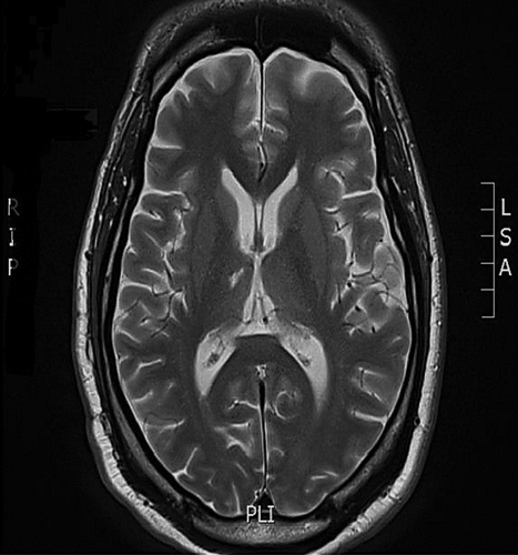
The patient is a man in his 40s, with community college education and no medical history, who developed vasculitis of unknown etiology involving the thalamoperforating vessels. Infarction largely involved the right thalamus (Figure 1).

Physical aggression was of new onset. The anterior nucleus is the principal relay nucleus for the limbic system, thought to play a role in emotion; it receives the mammilothalamic tract, which, in turn, projects to the cingulate gyrus.2 The dorsomedial nucleus (DM) similarly retains interconnections with the prefrontal cortex, which helps regulate affect and planning. Inputs to the DM include not only the prefrontal cortex but also elements of the limbic system, e.g., the amygdala. Successful treatment of aggression included divalproex sodium 1,000 mg p.o. od, citalopram 20 mg p.o. od, and olanzapine 5 mg p.o. od.
Previously unrecognized periods of diminished arousal were observed. These seemed to be of two types: In one, preceding verbal automatisms with focal motor activity were present. Despite a normal waking EEG, consideration to partial complex seizures was given. Seizures secondary to subcortical involvement have been reported,3 and the thalamus, including the reticular nucleus2 and the anterior nucleus,4 may play a role. Gabapentin was initiated at 300 mg p.o. tid, adjunctive to valproic acid. Residual periods of prolonged somnolence, often of quick onset, remained. Ascending projections from the reticular formation, which collects information from multiple sensory modalities, terminate in the thalamus, especially the intralaminar nuclei, and are involved in arousal.1 Modafinil 100 mg p.o. bid was started. A sleep study was conducted and detection of significant central apnea prompted use of continuous positive airway pressure (CPAP). Significantly greater alertness resulted with the above measures.
Cognitive changes were noted post-stroke. In particular, neuropsychological deficits were obvious in delayed memory (Repeatable Battery for the Assessment of Neuropsychological Status [RBANS] subtests List Learning and Story Memory), executive functioning (Stroop Color Word Test, Trails B), and visuospatial ability (RBANS subtest Figure Copy). Visuospatial deficits may be especially prominent in right-sided thalamic lesions,2 such as with this patient. The anterior nucleus of the thalamus maintains direct and indirect connections with the hippocampus, thought to subserve memory. Executive function is mediated in part by prefrontal cortex, as above, connected with the dorsomedial nucleus. Interestingly, although not explored in this case, there is a report of improvement in executive functioning in thalamic infarction with the use of donepezil.5
Damage to thalamic circuits results in wide-ranging effects, including arousal, cognition, and behavior. Knowledge of functional anatomy is important in the recognition and understanding of such impairment. Successful management may involve use of antiepileptics, antidepressants, antipsychotics, continuous positive airway pressure (CPAP), and stimulants.
1 : The Human Brain: An Introduction to Its Functional Anatomy, 4th Edition. Maryland Hgts., MO, Mosby, 1999Google Scholar
2 : Vascular syndromes of the thalamus. Stroke 2003; 34:2264–2278Crossref, Medline, Google Scholar
3 : The role of subcortical structures in human epilepsy. Epilepsy Behav 2002; 3:219–231Crossref, Medline, Google Scholar
4 :
5 . Effects of donepezil on behavioral manifestations of thalamic infarction: a single-case observation. Front Neurol 2:16Medline, Google Scholar


