Melancholia Associated With Severe Cognitive Disorders as the Expression of Late-Onset Postpartum Anti–N-Methyl-d-Aspartic Acid Receptor Limbic Encephalitis
To the Editor: Patients with anti–N-methyl-d-aspartic acid (NMDA) receptor encephalitis usually present with a neuropsychiatric disorder.1–3 Here we report the case of a young woman who presented with late-onset postpartum limbic encephalitis and benefited from neuropsychological follow-up until her cognitive recovery after 6 months.
Case Report
In January 2011, a 31-year-old woman suffering from paresthesia associated with melancholia was admitted to the hospital emergency department. Because a medical condition was apparently ruled out, the patient received a diagnosis of major depressive disorder with mood-congruent psychotic features and she was transferred to a psychiatry department. Results of her EEG, cerebral angiogram, CT scan of the head, and standard biological investigations were normal.
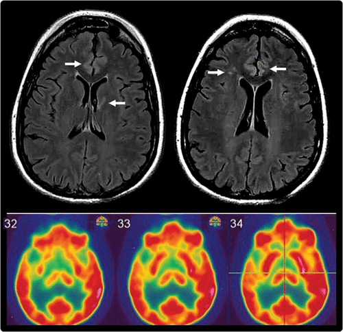
The patient was initially treated with mirtazapine 30 mg and clorazepate 60 mg daily. After 3 weeks, the patient was switched to clomipramine 200 mg and olanzapine 20 mg daily because of treatment inefficacy; however, she did not improve. Her psychiatric symptoms were accompanied by the following cognitive impairments: Mini-Mental State Examination score (25/30), temporal and spatial disorientation, and attentional and episodic memory impairments with relative preservation of the memory trace but encoding/retrieval impairment fitting with a dysexecutive syndrome (Frontal Assessment Battery score, 14 of 18; normal is >16). An EEG showed focal slow activity in the left temporal lobe. Am MRI scan of the brain revealed fluid-attenuated inversion recovery hyperintensities of the cingulate cortical ribbon bilaterally and in the white matter (Figure 1). Brain perfusion scintigraphy showed a bilateral hypoperfusion in the medial temporal lobe and in the right insular cortex. A positron emission tomography scan was performed on the same day, which detected hypometabolism in the right insular and temporal cortices. The CSF was positive for anti–NMDA receptor antibodies.

FIGURE 1. An MRI Scan With Fluid-Attenuated Inversion Recovery Contrast and a [18F]Fluorodeoxyglucose Positron Emission Tomography Scana
aThe upper image is an MRI scan with fluid-attenuated inversion recovery contrast showing the cingulate cortex and white matter hyperintensities (right frontal centrum semiovale and left genu of the internal capsule), whereas the lower image is an [18F]fluorodeoxyglucose positron emission tomography scan showing an extended hypometabolism in the right superior temporal gyrus, temporal pole, insula, and putamen (120×116 mm; 150×150 dots per inch).
The patient was diagnosed with anti–NMDA receptor limbic encephalitis. Treatment with corticoids (1 mg/kg) and twice-weekly immunoglobulin perfusions was initiated for 3 months, followed by one perfusion per month for a year. The patient fully recovered in 5 weeks and has shown no relapse over the past 2 years. Neuropsychological tests, conducted every 2 months, revealed that the patient’s impairments were restored after 6 months, which is delayed compared with other cases.4,5 A whole-body positron emission tomography scan and pelvic echography excluded the presence of a tumor.
Discussion
Anti–NMDA encephalopathy and other autoimmune diseases may present in the postpartum period6; however, in this case, the occurrence of illness was late (11 months after giving birth). According to the literature, the delayed diagnosis and the absence of a tumor seem to be pejorative factors for the response to treatment7 and frequently require many drug associations.4,5 However, in this case, all of the neuropsychiatric and cognitive symptoms were restored with only corticoid and immunoglobulin treatments.
Cognition is rarely properly evaluated,4,5 although it helped to orient the diagnosis by suggesting a focal dysfunction in our case. Moreover, its follow-up allowed us to substantiate subjective complaints, which were not obvious during the clinical evaluation, although they affected occupational and social outcomes. This may contribute to the “apparently” delayed recovery of this case compared with the literature, because “recovery” was defined by the normalization of cognitive functioning. Finally, in cases of persistent impairment, the neuropsychological evaluation might guide cognitive rehabilitation.
1 : Paraneoplastic encephalitis, psychiatric symptoms, and hypoventilation in ovarian teratoma. Ann Neurol 2005; 58:594–604Crossref, Medline, Google Scholar
2 : Paraneoplastic anti-N-methyl-D-aspartate receptor encephalitis associated with ovarian teratoma. Ann Neurol 2007; 61:25–36Crossref, Medline, Google Scholar
3 : Clinical experience and laboratory investigations in patients with anti-NMDAR encephalitis. Lancet Neurol 2011; 10:63–74Crossref, Medline, Google Scholar
4 : Anti-NMDA-receptor encephalitis presenting as postpartum psychosis in a young woman, treated with rituximab. Ann Saudi Med 2012; 32:421–423Crossref, Medline, Google Scholar
5 : Pregnancy outcome in anti-N-methyl-D-aspartate receptor encephalitis. Obstet Gynecol 2012; 120:480–483Crossref, Medline, Google Scholar
6 : Anti-NMDA receptor encephalitis. The disorder, the diagnosis and the immunobiology. Autoimmun Rev 2012; 11:863–872Crossref, Medline, Google Scholar
7 : Response of anti-NMDA receptor encephalitis without tumor to immunotherapy including rituximab. Neurology 2008; 71:1921–1923Crossref, Medline, Google Scholar


