Numbers Written Counterclockwise in the Clock Drawing Test
To the Editor: A recent review (Windows to the Brain: The Clock Drawing Task: Common Errors and Functional Neuroanatomy)1 described Rouleau’s qualitative system and the relationship between clock-drawing errors and brain networks. Frontoparietal and frontostriatal circuits are important for visuospatial and executive functions necessary for accurate clock-drawing. We highlight the error of counterclockwise numbers as spatial and/or planning deficits according to Rouleau’s system and argue that most of correctly sequenced numbers 1–12 written counterclockwise may suggest memory-system involvement.
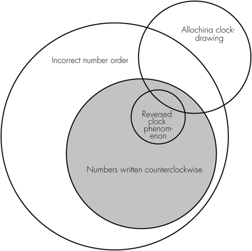
There are various number-order errors in the clock drawing test (CDT; Figure 1), and examples of representative clock-drawing appear in the supplement attached to this letter. Mendez suggested that >6 correctly sequenced of the same symbol type falls under the concept of “numerical factors,” which encode semantic and spatially-coded knowledge that a clock-face contains the numbers 1–12 arranged to represent hours.2

The reversed clock phenomenon (RCP) is an extreme example, in which 1–12 are written counterclockwise. Stroke patients with RCP often have lesions involving the right parietal lobe and subcortical area. Few stroke patients have parietotemporal lobe lesions, but lesions that involve multiple cortices make it difficult to clarify the role of the temporal lobes. RCP stroke patients show evidence of unilateral spatial neglect. Attentional orientation and visuospatial dysfunction, especially spatial representation, have been implicated in RCP stroke cases. Nevertheless, patients with Alzheimer’s disease (AD) and RCP did not show unilateral spatial neglect in Brugnolo’s study.3 The involvement of temporal lobes in AD patients with RCP supports the view that they are important for the visual-semantic retrieval of clock representation in AD patients.
The CDT can involve an instruction to draw a clock-face from memory (i.e. draw-to-command). Alternatively, a subject can be asked to copy a clock (i.e., copy-to-command), which places less demand on executive functions and semantic memory.1 RCP or counterclockwise writing by dementia patients was mostly in the clockwise order for the copy-to-command. Two subacute stroke patients and RCP with different lesions can produce similar clocks for the draw-to-command (i.e., allochiria), but different clocks for the draw-to-copy.4,5 We suggest that the combination of copy-to-command and draw-to-command provides more information about the availability of cognitive functions.
The CDT is simple and useful for monitoring dynamic cognitive change. A further understanding of the CDT results will help us design clinical rehabilitation programs to address specific cognitive impairments.
1 : The clock drawing task: common errors and functional neuroanatomy. J Neuropsychiatry Clin Neurosci 2012; 24:260–265Link, Google Scholar
2 : Development of scoring criteria for the clock drawing task in Alzheimer’s disease. J Am Geriatr Soc 1992; 40:1095–1099Crossref, Medline, Google Scholar
3 : The reversed clock drawing test phenomenon in Alzheimer’s disease: a perfusion SPECT study. Dement Geriatr Cogn Disord 2010; 29:1–10Crossref, Medline, Google Scholar
4 : On the different mechanisms of spatial transpositions: a case of representational allochiria in clock drawing. Neuropsychologia 2003; 41:1290–1295Crossref, Medline, Google Scholar
5 : Spatial transpositions across tasks and response modalities: exploring representational allochiria. Neurocase 2004; 10:386–392Crossref, Medline, Google Scholar


